

Overview
The urinary bladder is a hollow, distensible muscular organ that stores urine produced by the kidney and assists in micturition.

Location
- Anterior, midline, extraperitoneal pelvic organ
- Expands superiorly into the abdominal cavity when distended, otherwise lies entirely within the pelvic cavity
Gross anatomy
- Inverted pyramid, triangular in shape when empty
- Apex – directed towards the pubic symphysis, connected to umbilicus by the median umbilical ligament
- Base – triangular base facing posteroinferiorly
- Trigone – smooth triangular interior surface of the base, between openings of the ureters (above and laterally) and the internal urethral orifice (below and centrally).
- Ureteric orifices
- Angle of insertion of ureters causes a valve, closing with bladder distension

Surfaces
- Superior surface
- Covered by peritoneum, which continues posteriorly to cover floor of rectovesical pouch.
- Inferolateral surfaces (x2)
- Cradled by the anterior parts of the levator ani, the superior surface on which small bowel and sigmoid colon and uterus lie
- Lies against the anterior pelvic diaphragm and obturator internus
- Bladder neck
- Most inferior part of the bladder, where the base and inferolateral surfaces meet
- Surrounds the origin of the internal urethral orifice but doesn’t contribute to urinary continence, instead acts to prevent seminal regurgitation in male.
Arterial Supply
Superior and inferior vesical arteries (from the anterior division of internal iliac artery)
Venous Drainage
Vesical and prostatic venous plexus into internal iliac veins and internal vertebral veins
Lymphatics
- Initial lymphatic drainage from the bladder is primarily into the external iliac, obturator, internal iliac (hypogastric), and common iliac nodes.
- Following the drainage to these sentinel pelvic regions, spread may continue to the presacral, paracaval, interaortocaval, and para-aortic lymph node chains.
Innervation
- Sympathetic – hypogastric nerve (T12 – L2). It causes relaxation of the detrusor muscle, promoting urine retention.
- Parasympathetic – pelvic nerve (S2 – S4). Increased signals from this nerve causes contraction of the detrusor muscle, stimulating micturition.
- Somatic – pudendal nerve (S2 – 4). It innervates the external urethral sphincter, providing voluntary control over micturition.
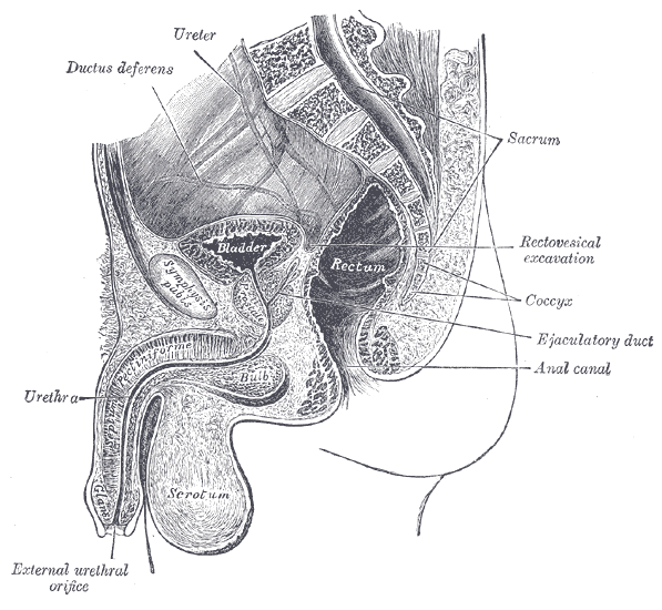
Relations
- Anterior: Symphysis pubis, retropubic space
- Posterior: Rectum and rectovesical pouch, seminal vesicles posteroinferiorly
- Inferior: Perineal membrane and deep perineal pouch, base of prostate (males), obturator internus
- Lateral: levator ani muscle, ischioanal fossa and anterior recesses
Variants
- Septated bladder – complete or incomplete, in frontal or sagittal plane
- Ureterocele – small distal dilatation forming sac-like pouch, often associated with a duplicated collection system
- Agenesis secondary to persistent cloaca – defect in which rectum, vagina (females) and urinary tract form single common channel
- Urachal cyst – cyst that develops in urachus, primitive structure that connects umbilical cord to bladder in developing foetus