

Overview
A pear-shaped, thick-walled, muscular secondary sex organ in the midline of the female pelvis, responsible for the transportation and maintenance of gametes.
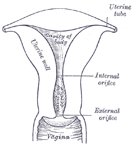
Structrue
Three internal layers:
- Endometrium
- Myometrium
- Perimetrium – serous layer of visceral peritoneum.
- Fundus – superior, rounded portion above the plane of the uterine tubes
- Body – midsection, surrounds the cavity
- Cavity – inverted triangle in coronal cross-section
- Cervix – directed posteroinferiorly, projects into anterior wall of vagina
- Uterine (Fallopian) tubes – project laterally from cornu of uterus on each side and has various segments (lateral to medial)
- Fimbriae – finger-like projections that drape over the ovary
- Infundibulum – a funnel-shaped lateral part that drapes over the ovary with the fimbriae emanating from it. Opens into the peritoneal cavity at the abdominal ostium
- Ampulla – the widest and longest section
- Isthmus – immediately lateral to the uterus, it is the narrowest segment
- Interstitial or intramural segment – the section within the myometrium
Position
Normally anteverted with respect to the vagina and antiflexed with respect to the cervix.
- Anteverted: Rotated forward, towards the anterior surface of the body.
- Anteflexed: Flexed, towards the anterior surface of the body.
Thus, the uterus normally lies immediately posterosuperior to the bladder, and anterior to the rectum.

Ligaments
- Broad ligament – A double layer of peritoneum attaching the sides of the uterus to the pelvis. It acts as a mesentery for the uterus and contributes to maintaining it in position. Contains:
- B: bundle (ovarian neurovascular bundle)
- R: round ligament
- O: ovarian ligament
- A: artifacts (vestigial structures)
- D: duct (oviduct) – a.k.a. the Fallopian or uterine tube.
- Broad ligament parts (inferior to superior):
- Mesometrium – Surrounds the uterus and runs laterally to cover the external iliac vessels. Encloses proximal part of round ligament
- Mesovarium – Associated with the ovaries. Projects from posterior surface of broad ligament to attach to anterior surface of ovaries, enclosing its neurovascular supply
- Mesosalpinx – Originates superior to mesovarium to enclose fallopian tubes.
- Round ligament – A remnant of the gubernaculum extending from the uterine horns to the labia majora via the inguinal canal. It functions to maintain the anteverted position of the uterus. It is continuous with the ovarian ligament laterally.
- Ovarian ligament – Joins the ovaries to the uterus.
- Cardinal ligament – Located at the base of the broad ligament, the cardinal ligament extends from the cervix to the lateral pelvic walls. It contains the uterine artery and vein in addition to providing support to the uterus.
- Uterosacral ligament – Extends from the cervix to the sacrum. It provides support to the uterus.

Blood supply
Uterus
Uterine artery, branch of the anterior division of internal iliac artery.
Fallopian Tube
Tubal branch of the ovarian artery (lateral one-third) and terminal (tubal) branch of the uterine artery (medial two-thirds)
Venous drainage
Uterus
Venous drainage is via a plexus in the broad ligament that drains into the uterine veins, which empty into the internal iliac vein.
Fallopian Tube
- Lateral one-third via the pampiniform plexus to the ovarian veins (right drains into the inferior vena cava, left drains into left renal vein)
- Medial two-thirds via the uterine plexus to the internal iliac vein
Lymphatic drainage
Ovaries and uterine tubes
Lymphatic drainage from the ovaries and uterine tubes follow the ovarian veins directly to the lumbar lymph nodes at L1-2 (i.e., the caval and aortic lymph nodes), bypassing the iliac nodes (similar to the testis and epididymis).
Fundus and upper body
- Right lumbar lymph nodes – composed of precaval, retrocaval, lateral lymph nodes
- Left lumbar lymph nodes – composed of preaortic, retroaortic, lateral aortic lymph nodes
- Pass laterally in upper broad ligament to join lymphatics from uterine tube and ovaries before running over pelvic brim with ovarian vessels
- Some drain along round ligament into superficial inguinal nodes, through the inguinal canal
Lower body and cervix
Lymphatic drainage runs laterally in lower parts of the broad ligament and demonstrate 3 pathways:
- External Iliac Nodes (along pelvic brim)
- Passage through medial external iliac vein, then middle and lateral before ascending into common iliac chain
- Internal Iliac Nodes
- Follows branches of internal iliac artery before ascending to common iliac nodes
- Presacral route
- Lymphatic plexus anterior to sacrum and coccyx before ascending to common iliac nodes
Innervation
- Sympathetic: uterovaginal plexus
- Parasympathetic: pelvic splanchnic nerves (S2-S4)
- The cervix is largely innervated by the inferior nerve fibres of the uterovaginal plexus.
- The afferent fibres mostly ascend through the inferior hypogastric plexus to enter the spinal cord via T10-T12 and L1 nerve fibres.

Relations
• Anterior: bladder, vesicouterine pouch
• Posterior: rectum, rectouterine pouch (of Douglas)
• Lateral: broad ligament, uterine vessels, uterine tubes
• Inferior: bladder, communicates with vagina
Variant anatomy
Uterus
- Uterus didelphys
- Septated uterus
- Bicornuate uterus
- Unicornuate uterus
- Hypoplasia or agenesis of vagina, cervix, fundus or tubes
Uterine artery
- Arises from the umbilical artery directly or via common stem
- Arises from the inferior gluteal directly or via common stem
- Variable origin along internal iliac: 1st branch (19%), 2nd or third branches (71%)
- Passes ureter posteriorly (5%)
- Bifurcation or trifurcation at uterus
Fallopian tubes
- absent unilateral
- hypoplastic unilateral
- hypoplastic bilateral
- abnormal entry into uterine body or fundus