

Overview
An oblong-shaped, glandular retroperitoneal organ (except the tail), located within the anterior pararenal space at the level of the transpyloric plane (level of L1). The head and uncinate process lie just right to the midline and extends to the left hypochondrium. It has both digestive (exocrine) and hormonal (endocrine) functions.
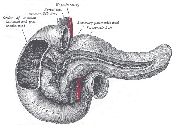

Structure
- Main pancreatic duct
- Dorsal duct proximal to dorsal-ventral fusion
- Continuous with Duct of Wirsung (ventral duct distal to dorsal ventral fusion)
- Pancreatic duct of Wirsung – unites with CBD and drains into major papilla into D2 of duodenum
- Accessory Pancreatic Duct (of Santorini) – dorsal duct distal to dorsal-ventral fusion, drains anterior and superior aspect of the head
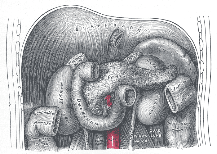
Arterial Supply
- Gastroduodenal artery (branch of common hepatic artery)
- Anterior and posterior superior pancreaticoduodenal artery (branch of gastroduodenal artery)
- Dorsal pancreatic (branch of splenic artery)
- Continues as inferior pancreatic artery
- Greater pancreatic artery (branch of splenic and inferior pancreatic artery)
- Anterior and posteior inferior pancreaticoduodenal artery (branch of superior mesenteric artery)
Venous Drainage
- Anterior and posterior superior pancreaticoduodenal vein – drains into portal vein
- Anterior and posterior inferior pancreaticoduodenal vein – drains to superior mesenteric vein
- Splenic vein – drains into portal vein
Lymphatic Drainage
- Vessels follow the arteries, drain into pre-aortic, pancreaticosplenic and coeliac nodes
Innervation
- Distributed with arteries via coeliac plexus
- Sympathetic – Greater splanchnic nerve (T6-10)
- Parasympathetic – Vagal trunks
Relations
Head
- Anterior: transverse colon, stomach
- Posterior: Splenic vein, superior mesenteric artery and vein, abdominal aorta, inferior vena cava, left renal artery and vein
- Superior: epiploic foramen
Neck
- Posterior: Superior mesenteric vein and portal vein
Body
- Runs across renal vein, abdominal aorta, left crus of diaphragm, left psoas major muscle, lower pole of left adrenal gland and left kidney
Tail
Development
- It is of endodermal origin.
- Development begins with the formation of a dorsal and ventral pancreatic bud that arises from the duodenal part of the foregut
- Each joins with the foregut through a duct.
- The dorsal pancreatic bud forms the neck, body, and tail of the developed pancreas
- The ventral pancreatic bud forms the head and uncinate process
Variants
Pancreas
- Pancreas divisum
- Most common variant
- Failure of fusion of dorsal and ventral pancreatic anlages, resulting in the dorsal pancreatic ducting draining most of the parenchyma via the minor papilla.
- Annular pancreas
- Bifid pancreatic tail
- Accessory or ectopic pancreas
- Meandering main pancreatic duct
- Ansa pancreatica (rare) – Communication between main and accessory pancreatic duct
- Anomalous pancreaticobiliary junction – union of the pancreatic duct and common bile duct outside the duodenal wall
Pancreatic Duct
- Double accessory pancreatic duct (Santorini)
- Double main pancreatic duct (Wirsung)
- Anastomosis between ducts
- Crossing or double-crossing of ducts
- No communication between ducts
- Tortuosity of ducts
- Absence of accessory pancreatic duct