

Overview
The psoas major is a long, thick, fusiform muscle located in the lumbar region of the posterior abdominal wall, in the gutter between the vertebral bodies and transverse processes.
Origin
The psoas major muscle has an extensive and continuous origin, spanning the vertebral bodies and intervertebral discs of T12 (lower border of body) – L4/5 (upper border of body) superficially, and the transverse processes of L1-L5 vertebrae deep.
Course
- The muscle extends inferolaterally through the true pelvis towards the thigh
- The lateral most fibres fuse with the iliacus muscle to form the iliopsoas muscle at the level of L5 – S2
- The iliopsoas muscle then passes inferiorly, deep to the inguinal ligament and anterior to the joint capsule of the hip
Insertion
Lesser trochanter of the femur as the iliopsoas tendon
Coverings
Psoas fascia – the psoas muscle is enclosed by the psoas fascia (which retains pus in psoas abscesses), part of the iliac fascia, which invests the surface of the muscle. The fascia is attached to the vertebral bodies, fibrous arches, and the transverse processes, and extends along the pelvic brim attached to the iliopubic eminence at the margins of the muscle. The lateral edge blends with the anterior layer of the lumbar fascia (over quadratus lumborum).
Action
The action of this muscle is complex, acting to both laterally flex the lumbar spine as well as stabilize and flex the thigh. The antagonist is the gluteus maximus.
Relations
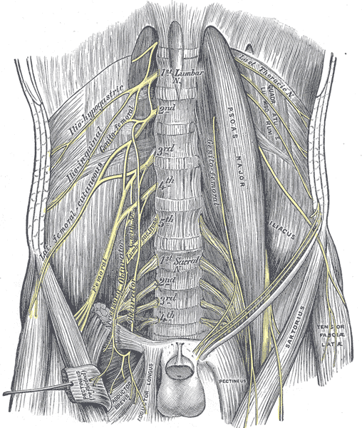
The lumbar plexus is embedded within the muscle and its branches emerge from it:
- Anterior aspect: genitofemoral nerve
- Lateral border: iliohypogastric, ilioinguinal, lateral femoral cutaneous and femoral nerves
- Medial border: obturator nerve and lumbosacral trunk
Superiorly
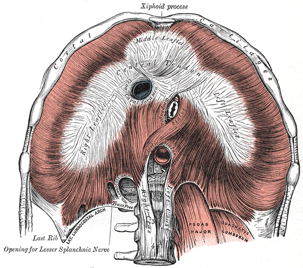
- Diaphragm (anteriorly)
Laterally
- Quadratus lumborum
- Femoral nerve
Medially
- Inferior vena cava (right) and abdominal aorta (left)
- Sympathethic trunk
- Para-aortic lymph nodes
- Pectineus and the femoral vein
Anteriorly
- Retroperitoneum and retroperitoneal structures
- Kidneys
- Ureters
- Gonadal vessels
- In the thigh, psoas major contributes to the floor of the femoral triangle.
- Iliopsoas tendon lies deep to the fascia lata, sartorius, rectus femoris and deep femoral artery.
Posteriorly
- Separated from the joint capsule of the hip by the iliac bursa
- Part of the external vertebral venous plexus (in front of the transverse process)
- Four lumbar arteries and veins pass beneath the four arches and run laterally behind the psoas accompanied by the sympathetic rami
Lumbosacral triangle of Marcille
- Contents from medial to lateral: obturator nerve, iliolumbar artery, lumbosacral trunk, sympathetic trunk
Innervation
The psoas muscle is innervated by the anterior branches of spinal nerves L1 – L3 (mainly L2) from the lumbar plexus
Arterial Supply
- Lumbar branches (of the aorta) – supplies the upper part
- Iliolumbar branch (branch of the posterior division of the internal iliac artery) – supplies the middle part
- Obturator branch (of the anterior division of the internal iliac artery)
- Branches of the external iliac artery and femoral arteries also contribute to the supply of the lower part
Venous Drainage
- Venous drainage follows arterial supply, draining into the femoral, external iliac and internal iliac veins as well as directly into the inferior vena cava
- Lumbar veins
- First and second lumbar veins to azygos system
- Third and fourth to inferior vena cava
Lymphatic Drainage
- Follows the lumbar vessels into cisterna chyli
Variant Anatomy
- Asymmetry of the psoas major muscle, which is usually not clinically significant
- Associated with psoas minor – origin from T12 and L1 vertebrae and inserted on the arcuate line and iliopubic eminence
- Accessory muscle slips – femoral nerve may or may not be split by accessory slip