

Overview
The musculature of the thigh is divided into anterior, medial and posterior compartments, each with distinct innervation and function.
The medial compartment of the thigh is composed of 5 muscles comprising the hip adductor group. These muscles extend from the anteroinferior external surface of the bony pelvis to the shaft of femur and proximal tibia. The majority of hip adductors are innervated by the obturator nerve (L2-L4) and supplied by blood mainly via the branches of the femoral and obturator arteries. It’s primary action is to produce adduction of the thigh at the hip joint. Additionally, they contribute to the stabilisation and balance of the pelvis and body posture while standing, walking, or running.
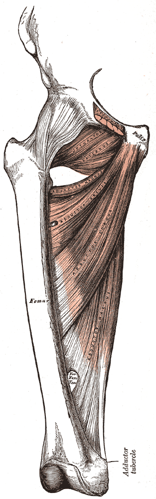
Adductor Magnus
The adductor magnus is the largest muscle in the medial compartment and lies posteriorly. Functionally, the muscle can be divided into two parts; the adductor part (adductor minimus – smaller) and the hamstring part (larger). The distal hamstring part features an adductor hiatus between the adductor magnus and femur, in which allows passage of the femoral artery and femoral vein (which then becomes the popliteal artery and popliteal vein distally).
- Origin & Insertion:
- Actions: Adduction of the thigh. The adductor component also flexes the thigh, with the hamstring portion extending the thigh.
- Innervation:
- Adductor part – obturator nerve (L2, L3, L4), branch of the lumbar plexus
- Hamstring part – tibial component of the sciatic nerve (L4-S3), branch of the sacral plexus
Adductor Longus
The adductor longus is a large, flat muscle partially covering the adductor brevis and magnus. It forms the medial border of the femoral triangle.
- Origin: Pubis
- Insertion: Fans out broadly, attaching to the linea aspera of the femur
- Actions: Adduction of the thigh
- Innervation: Obturator nerve (L2, L3, L4), branch of the lumbar plexus
Adductor Brevis
The adductor brevis is a short muscle, lying underneath the adductor longus. It lies in between the anterior and posterior divisions of the obturator nerve.
- Origin: Body of pubis and inferior pubic rami
- Insertion: Linea aspera on the posterior surface of the femur, proximal to the adductor longus.
- Actions: Adduction of the thigh
- Innervation: Obturator nerve (L2, L3, L4), branch of the lumbar plexus
Obturator Externus
The smaller muscles of the medial thigh located most superiorly.
- Origin: Membrane of the obturator foramen.
- Insertion: Passes under the neck of femur, attaching to the posterior aspect of the greater trochanter.
- Actions: Adduction and lateral rotation of the thigh.
- Innervation: Obturator nerve (L2, L3, L4), branch of the lumbar plexus
Gracilis
The most superficial and medial of the muscles in this compartment. It crosses at both the hip and knee joints.
- Origin: Inferior rami of the pubis and the body of the pubis.
- Insertion: Descending almost vertically down the leg and attaches to the medial surface of the tibia, between the tendons of the sartorius (anteriorly) and the semitendinosus (posteriorly).
- Actions: Adduction of the thigh at the hip, and flexion of the leg at the knee.
- Innervation: Obturator nerve (L2, L3, L4), branch of the lumbar plexus
Anatomical Variants
Obturator nerve
- Obturator nerve division in pelvis
- Obturator nerve division within obturator canal
- Obturator nerve division within thigh
- Posterior branch supplying adductor brevis