

Overview
The musculature of the lower leg is divided into anterior, lateral (fibular), posterior (superficial and deep) compartments, defined by intermuscular septa. The two muscles of the lateral compartment of the leg acts to everse the foot.
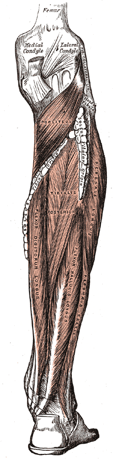
Fibularis Longus
The fibularis longus is the larger and more superficial muscle
- Origin: Superior and lateral surface of the fibula and the lateral tibial condyle.
- Course:
- The fibres converge into a tendon which runs posterior to the lateral malleolus
- It then passes under the superior and inferior fibular retinaculum together with the tendon of the fibularis brevis, but wraps under the foot
- It extends obliquely forward on the plantar surface of the foot
- Insertion: Lateral aspect of the medial cuneiform and lateral base of the first metatarsal
- Actions: Eversion and plantarflexion of the foot. Also supports the lateral and transverse arches of the foot.
- Innervation: Superficial fibular (peroneal) nerve, L4-S1.

Fibularis Brevis
The fibularis brevis muscles is deeper and shorter than the fibularis longus.
- Origin: Inferolateral surface of the fibular shaft
- Course:
- The muscle belly forms a tendon, which descends with the fibularis longus into the foot.
- It travels posteriorly to the lateral malleolus, passing under the superior and inferior fibular retinaculum together with the tendon of the fibularis longus
- It then passes over the calcaneus and the cuboidal bones.
- Insertion: Tubercle on the fifth metatarsal
- Actions: Eversion of the foot
- Innervation: Superficial fibular (peroneal) nerve, L4-S1

