

Overview
The musculature of the lower leg is divided into anterior, lateral (fibular), posterior (superficial and deep) compartments, defined by intermuscular septa. The posterior compartment of the leg contains seven muscles, organised into two layers, separated by a band of fascia – superficial and deep. The posterior leg is the largest of the three compartments, and collectively, the muscles in this area plantarflex and invert the foot.

Superficial Muscles
The superficial muscles form the calf shape of the posterior leg.
Gastrocnemius
The gastrocnemius is the most superficial and has a medial and lateral head, which converge to form a single muscle belly.
- Origin:
- Lateral head – lateral femoral condyle
- Medial head – medial femoral condyle
- Insertion: The fibres of the medial and lateral head converge to form a single muscle belly, which combines with the soleus to from the calcaneal tendon. The calcaneal tendon inserts onto the calcaneal tuberosity on the calcaneus.
- Actions:
- Plantarflexes the foot at the ankle joint
- Flexes at the knee
- Innervation: Tibial nerve, terminal branch of the sciatic nerve (L4-S3)
Plantaris
The plantaris is a small muscle with a long tendon, which can be mistaken for a nerve as it descends down the leg.
- Origin: Lateral supracondylar line of the femur
- Course: Descends medially, condensing into a long tendon that runs down the leg, between the gastrocnemius and soleus.
- Insertion: The tendon blends with the calcaneal tendon to insert onto the calcaneal tuberosity on the calcaneus
- Actions:
- Plantarflexes the foot at the ankle joint
- Flexes at the knee
- Innervation: Tibial nerve, terminal branch of the sciatic nerve (L4-S3)
Soleus
A large flat muscles located deep to the gastrocnemius, so named due to its resemblance of a flat fish called a sole.
- Origin: Soleal line of the tibia and proximal fibular area.
- Insertion: The muscle narrows in the lower part of the leg, combining with the gastronemius distally to form the calcaneal tendon, to insert onto the calcaneal tuberosity on the calacenus
- Actions: Plantarflexes the foot at the ankle joint
- Innervation: Tibial nerve, terminal branch of the sciatic nerve (L4-S3)
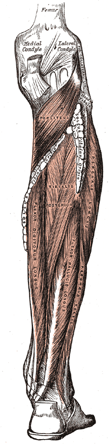
Deep Muscles
There are four muscles in the deep compartment of the posterior leg.
Popliteus
The popliteus is located superiorly in the leg, behind the knee joint, forming the base of the popliteal fossa. Acts only on the knee joint.
- Origin: Lateral condyle of the femur and the posterior horn of the lateral meniscus
- Course: It runs inferomedially towards the tibia
- Incidence: Inserts above the origin of the soleus muscle
- Actions: Laterally rotates the femur on the tibia – ‘unlocking’ the knee joint so that flexion can occur
- Innervation: Tibial nerve, terminal branch of the sciatic nerve (L4-S3)
- Relations:
- Posterior: Popliteus bursa (between popliteal tendon and posterior surface of the knee joint)
Tibialis Posterior
The tibialis posterior is the deepest out of the four muscles. It lies between the flexor digitorum longus and the flexor hallucis longus.
- Origin: Interosseous membrane between the tibia and fibula, and posterior surfaces of the two bones
- Insertion: The tendon enters the foot posterior to the medial malleolus via the tarsal tunnel, and attaches to the plantar surfaces of the medial tarsal bones.
- Actions: Inverts and plantarflexes the foot, maintains the medial arch of the foot.
- Innervation: Tibial nerve, terminal branch of the sciatic nerve (L4-S3)
- Relations: Posterior tibial artery and tibial nerve run along it’s superficial surface

Flexor Digitorum Longus
The FDL is a smaller muscle than the flexor hallucis longus. It is located medially in the posterior leg.
- Origin: Medial surface of the tibia
- Course:
- Passes behind medial malleolus, separated from tibialis posterior by a fibrous septum
- Crosses tibialis posterior above the medial malleolus
- Passes obliquely forward and lateralward, superficial to the deltoid ligament of the ankle joint into the sole of the foot
- Crosses flexor hallucis longus at the level of the navicular bone, receives a strong tendinous slip from it
- Expands, joined by quadratus plantae and divides into 4 tendons
- Insertion: Attaches to the plantar surfaces of the lateral four digits
- Actions: Flexes the lateral four toes.
- Innervation: Tibial nerve, terminal branch of the sciatic nerve (L4-S3)
Flexor Hallucis Longus
The flexor hallucis longus muscle is found on the lateral side of leg.
- Origin: Originates from the posterior surface of the fibula
- Course:
- Fibres pass down and back, through the tarsal tunnel on the medial side of the foot
- Lies in a groove crossing the posterior surface of the lower end of tibia, between medial and lateral tubercles of the posterior surface of talus and undersurface of sustentaculum tali
- The groove is lined by a mucous sheath
- Runs between the two heads of flexor hallucis bevis
- Insertion: Attaches to the plantar surface of the phalanx of the great toe
- Actions: Flexes the great toe
- Innervation: Tibial nerve, terminal branch of the sciatic nerve (L4-S3)
- Relations: Covers the distal course of the fibular artery
Bursa
Two bursae associated with the calcaneal tendon minimise friction during movement:
- Subcutaneous calcaneal bursa – lies between the skin and the calcaneal tendon.
- Deep bursa of the calcaneal tendon – lies between the tendon and the calcaneus.
Anatomical Variants
- Absent plantaris (10% of people)