

Overview
The popliteal fossa is a diamond-shaped shallow depression located posterior to the knee joint that allows dynamic movement of neurovascular structures traversing from the thigh to leg during knee flexion and extension.
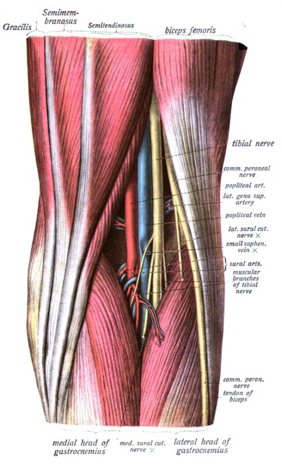
Borders
- Superomedial border – Lateral border of the semimembranosus muscle and tendon of semitendinosus superficially
- Superolateral border – Medial border of biceps femoris (short and long heads)
- Inferomedial border – Lateral border of the medial head of the gastrocnemius.
- Inferolateral border – Medial border of the lateral head of the gastrocnemius and the plantaris (deep to the gastrocnemius)
- Floor – superior to inferior:
- Popliteal surface of the femur
- Knee joint capsule
- Popliteus and fascia encasing the popliteus
- Roof – consists of two layers: popliteal fascia and skin. The popliteal fascia is continuous with the fascia lata of the leg.
Contents
- Fat (main component)
- Popliteal lymph nodes and vessels which follow the femoral vessels, draining into the deep inguinal nodes.
- Embedded in the fat.
- Beneath the popliteal fascia
- Along the popliteal vessels

Vascular Structures
- Popliteal artery
- Deepest structure. Distal continuation of the femoral artery after passing the adductor hiatus of the adductor magnus. Terminates at the inferior border of the popliteus muscle as the anterior tibial and common trunk of the posterior tibial artery
- Gives off five genicular branches which form a collateral network between the descending genicular artery of the femoral artery and popliteal artery: superior lateral, superior medial, middle, inferior lateral, and inferior medial genicular arteries.
- Popliteal vein
- At all levels, it lies in between the popliteal artery and tibial nerve
- Receives venous blood from anterior tibial vein, posterior tibial vein and peroneal vein
- Extends superiorly through the adductor hiatus before becoming the femoral vein
- Small saphenous vein
- Ascends from the lateral marginal vein of the foot and pierces the roof of the popliteal fossa to enter the popliteal vein in the lower half of the fossa popliteal lymph nodes
Nerves
The sciatic nerve branches into the tibial and common fibular (peroneal) nerves at the superior angle of the popliteal fossa and are the most superficial of the structures within the fossa:
- Tibial nerve (L4 – S3)
- Most superficial, larger than the common fibular nerve
- Enters the posterior leg between the heads of the gastrocnemius
- Gives off muscular, articular, and cutaneous (sural) branches
- Muscular branches in the distal portion of the fossa supply the medial and lateral heads of the gastrocnemius, soleus, plantaris, and popliteal muscles
- The sural nerve extends from the middle of the popliteal fossa to pierce the roof, providing innervation to the lower posterior half of the leg and lateral foot
- Common fibular (peroneal) nerve (L4 – S2)
- Courses to the lateral border where it wraps around the fibular head
- Gives off a communicating sural nerve which descends to pierce the roof in the lateral fossa and join the sural nerve
- Posterior femoral cutaneous nerve: descends and pierces the roof
Development
- Limb bud development of the lower extremity begins in the fourth week of gestation.
- Continues to lengthen into week 5 and undergoes a series of rotations and regional morphological development into the thigh, leg, and foot regions
Variants
- Aplastic or hypoplastic posterior tibial artery
- Hypoplastic or aplastic anterior tibial artery
- Trifurcation
- High origin of the anterior tibial artery
- High origin of the posterior tibial artery
- Anterior tibioperoneal trunk.
- High division of the sciatic nerve into tibial and common peroneal nerve