

Overview
The veins of the lower limb drain deoxygenated blood and return it to the heart. They can be divided into two groups – deep and superficial.
Deep veins are located underneath the deep fascia of the lower limb, accompanying the major arteries and following its naming convention. Superficial veins are found in the subcutaneous tissue, but eventually drain into the deep veins.
Deep Venous System
The deep venous drainage system of the lower limb is located beneath the deep fascia of the lower limb. As a general rule, the deep veins accompany and share the name of the major arteries in the lower limb. Often, the artery and vein are located within the same vascular sheath – so that the arterial pulsations aid the venous return.

Foot
Venous drainage of the foot is achieved by plantar veins, which drain the underside of the foot and dorsal veins, which drain the upper surface of the foot
- Digital veins – At the toes, interconnected venous plexuses of the plantar and dorsal regions form plantar and dorsal digital veins, respectively.
- Metatarsal veins – The plantar and dorsal digital veins course proximally in the metatarsal space, to form four plantar and dorsal metatarsal veins.
- Deep plantar venous arch – The plantar metatarsal veins course proximally within the intermetatarsal spaces to form the deep plantar venous arch. The deep plantar venous arch runs from the proximal end of the first interosseous space to the base of the fifth metatarsal and accompanies the deep plantar arterial arch
- Plantar veins – The deep plantar venous arch continues proximally medially as the medial plantar vein and laterally as the lateral plantar vein
- The medial plantar vein is usually doubled and runs along the medial border of the sole from the medial end of the deep plantar arch to the medial malleolus
- The lateral plantar vein lies between two muscle layers of the sole of the foot (quadratus plantae and abductor allucis)
- The confluence of the medial and lateral plantar veins at the medial malleolus forms the posterior tibial and the fibular (peroneal) veins
Leg
These veins combine to form the posterior tibial and fibular veins.
- The posterior tibial vein accompanies the posterior tibial artery, entering the leg posteriorly to the medial malleolus via the tarsal tunnel.
- It ascends in the deep posterior compartment of the lower leg, between the tibialis posterior and the soleus
- It emerges from the deep posterior compartment via the tendinous arch of soleus, to enter the inferior aspect of the popliteal fossa
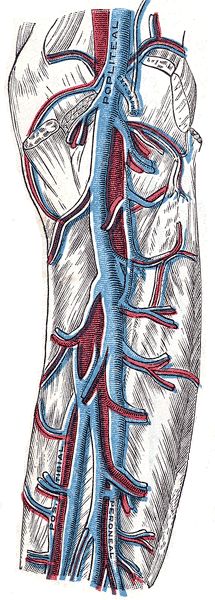
Knee
- Within the popliteal fossa, the anterior tibial, posterior tibial and fibular veins unite to form the popliteal vein.
- At the centre of the popliteal fossa, the popliteal vein is crossed by the tibial nerve (which crosses over it from medial to lateral)
- The popliteal vein enters the thigh via the adductor canal, together with the popliteal artery and popliteal vein.
Thigh
- Once the popliteal vein has entered the thigh, it is known as the femoral vein. It is situated anteriorly, accompanying the femoral artery.
- The deep vein of the thigh (profunda femoris vein) is the other main venous structure in the thigh. Via perforating veins, it drains blood from the thigh muscles. It then empties into the distal section of the femoral vein.
- The femoral vein leaves the thigh by running underneath the inguinal ligament, at which point it is known as the external iliac vein.
Gluteal Region
The gluteal region is drained by inferior and superior gluteal veins which empty into the internal iliac vein.
Superficial Venous System
Foot
- Metarsal veins – Dorsal metatarsal veins form the dorsal venous arch which lies over the proximal ends of the metatarsal bones
- Dorsal venous arch – The dorsal venous arch continues proximally as the medial and lateral marginal veins of the foot which receives various perforating branches on its course proximally, including the dorsal vein of the great toe
- Marginal veins
- The medial marginal vein continues to become the great saphenous vein
- The lateral marginal veins continues to become the small saphenous vein

Great Saphenous Vein
- Origin: Arises anterior to the medial malleolus as the continuation of the medial margin vein, formed by the dorsal venous arch of the foot and the dorsal vein of the great toe
- Course:
- It ascends up the medial side of the leg within the saphenous compartment of the subcutaneous tissue.
- Saphenous compartment – The saphenous compartment is bound posteriorly by the deep fascia and anteriorly by the saphenous fascia
- It first passes anterior to the medial malleolus at the ankle and ascends posterior to the medial condyle at the knee.
- At the upper thigh, it pierces the saphenous hiatus of the deep fascia of the thigh, 1-3 cm distal to the inguinal ligament, to drain into the femoral vein at the saphenofemoral junction in the femoral triangle.
- There is a venous valve 1-2 mm distal to the saphenofemoral junction
- It ascends up the medial side of the leg within the saphenous compartment of the subcutaneous tissue.

- Tributaries:
- Small saphenous vein (SSV): communicating branches in the region of the popliteal fossa
- Lateral accessory vein
- Superficial epigastric vein
- Superficial circumflex iliac vein
- Superficial external iliac vein
- Superficial external pudendal vein
- Deep external iliac vein
- Anterior accessory saphenous vein
- Termination: The great saphenous vein terminates by draining into the femoral vein immediately inferior to the inguinal ligament.
- Relations:
- Below the knee, the branches of the saphenous nerve are located posteriorly and anteriorly
- Above the knee the saphenous nerve is not closely related (i.e. is not within the saphenous compartment)
- Superficial medial lymphatic vessels follow the course of the greater saphenous vein, draining into the subinguinal group of inguinal lymph nodes

Small Saphenous Vein
- Origin: Arises posterior to the lateral malleolus as the continuation of the lateral marginal vein of the foot, formed by the dorsal venous arch of the foot, and the dorsal vein of the little toe
- Course:
- It moves up the posterior side of the leg, passing posteriorly to the lateral malleolus, along the lateral border of the calcaneal tendon.
- It moves between the two heads of the gastrocnemius muscle and empties into the popliteal vein in the popliteal fossa.
- Tributaries:
- Communicating branches with the great saphenous vein
- Cutaneous venous tributaries
- Communications:
- Communicates with deep venous system via numerous perforating veins
- Relations
- Accompanied by the sural nerve (branch of the tibial nerve)
- Superficial lateral lymphatic vessels follow the course of the greater saphenous vein, draining into the popliteal lymph nodes
Variants
Great Saphenous Vein
- Segmental hypoplasia or aplasia
- Duplication: only in the thigh
- Duplicated great saphenous vein lies within the saphenous compartment
- Enters common femoral together or separately
- Accessory saphenous veins: ascend with the SSV or GSV
- Lie outside the saphenous compartment
- Drains into superficial femoral vein
Small Saphenous Vein
- Duplication (~3.5%)
- Variable termination (i.e. not into the popliteal vein in the popliteal fossa)
- Vein of Giacomini – thigh extension of small saphenous vein. Courses between biceps femoris and semimembranosus muscles and drains into superficial, perforating or deep veins (~80%)
- Variable presence of communicating branches with the popliteal vein
- No connection to popliteal vein, drains variably into GSV
- Accessory saphenous veins
- Accessory saphenous vein may drain into common femoral vein (Mickey mouse sign on USS)