

Overview
The femoral vein is a large vein located deep within the thigh and is responsible for draining the lower limb. The femoral vein typically has four to five bicuspid valves at various points along its length.
Origin
Forms as the proximal continuation of the popliteal vein at the adductor opening (opening in the hamstring part of the adductor magnus)
Course
- The femoral vein then ascends in the adductor canal, initially posterolateral to the superficial femoral artery
- Adductor canal (Hunter’s canal, subsartorial canal) – a 15 cm long narrow conical tunnel extending from the apex of the femoral triangle to the adductor hiatus of the adductor magnus
- As the femoral vein ascends through the femoral triangle towards the base, it becomes medial to the femoral artery within the femoral sheath, lateral to the femoral canal
- It occupies the middle compartment of the femoral sheath
- Proximal to the confluence with the deep femoral vein, the femoral vein is known as the common femoral vein.
Termination
Terminates at the inferior margin of the inguinal ligament, where it ascends posterior to the ligament to become the external iliac vein.

Tributaries
- Numerous muscular tributaries
- Deep femoral vein (profunda femoris) vein
- Joins the femoral vein posteriorly 4-12 cm distal to the inguinal ligament
- formed by perforating veins that drain the surrounding muscle
- Great (long) saphenous vein
- Joins anteriorly or anteromedially
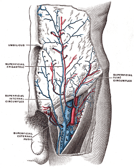
- Prior to joining the femoral vein at its proximal end, the great saphenous vein receives the external pudendal, superficial circumflex iliac, superficial epigastric and accessory saphenous veins,
- Lateral and medial circumflex femoral veins
Anatomical Variants
- Bifid or duplicated femoral veins