

General
Broad musculotendinous sheet shaped as upward-curved double domes attached at its circumference to the internal wall of the torso, at the inferior thoracic aperture. It separates the thoracic and abdominal cavities, and alters the volume of the thoracic cavity in inspiration and expiration by contracting and relaxing. At rest, the right dome lies slightly higher than the left due to the presence of the liver inferiorly.
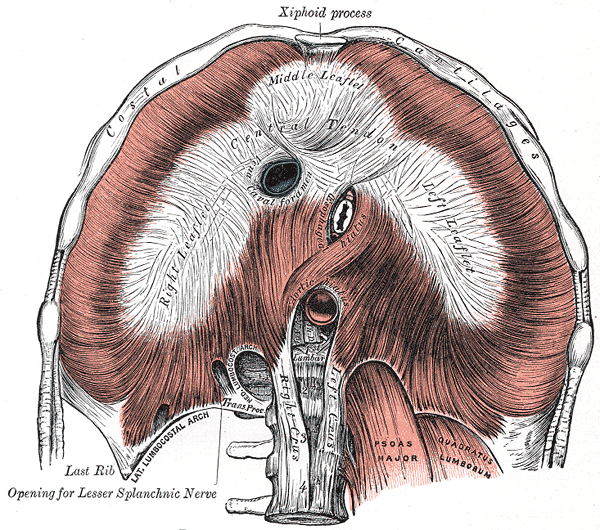
Attachments
Peripheral Attachments
- Lumbar vertebrae
- Ribs 7 – 10 indirectly via costal cartilage
- Ribs 11 – 12 directly
- Xiphoid process of the sternum
- Lateral Arcuate Ligament – from tip of rib 12 to transverse process of L1
- Medial Arcuate Ligament – from transverse process of L1 to anterior surface of vertebral bodies as left and right crura (tendinous attachment that blends with anterior longitudinal ligament)
Central Attachments
The cura (latin: leg) of the diaphragm are tendinous structures that extens below the diaphragm to the vertebral column. Together they form a tether for muscular structure.
- Right crus – Arises from vertebral bodies L1-3 and their intervertebral discs.
- Some fibres from the right crus surround the oseophageal hiatus, acting as a physiological sphincter to prevent gastric content reflux into the oesophagus.
- Larger and longer than the left
- Left crus – Arises from vertebral bodies L1-2 and their interverebral discs
- Median Arcuate Ligament – medial fibres of left and right crura meet in the midline to form median arcuate ligament. The area behind this is known as the aortic hiatus.
- Central tendon – the muscle fibres of the diaphragm combine to form a central tendon. This tendon ascends to fuse with the inferior surface of the fibrous pericardium. Either side of the pericardium, the diaphragm ascends to form left and right domes.
Apertures
Major Diaphragramtic Apertures
- T8 – Caval hiatus through the central tendon
- Transmits the inferior vena cava and right phrenic nerve
- T10 – Oesophageal hiatus left of the midline
- Transmits the oseophagus, anterior and posterior vagal trunks, oesophageal branch of left gastric artery and vein
- T12 – Aortic hiatus midline between the left and right crura
- Transmits the abdominal aorta, thoracic duct and azygous vein
Lesser Diaphragmatic Apertures
- Right crus of diaphragm (x 2)
- Transmits the right greater and lesser splanchnic nerves, occasionally the azygous vein.
- Left crus of diaphragm (x 3)
- Transmits the left greater and lesser splanchnic nerves and hemi-azygous vein
- Medial arcuate ligament
- Transmits the sympathethic trunk
- Lateral arcuate ligament
- Subcostal nerve and vessels
- Left phrenic nerve pierces the dome of the left hemidiaphragm
Sternocostal Foramina
- Sternocostal triangle (a.k.a. foramina of Morgagni or Larrey’s space) are small zones between the costal and sternal attachments of the diaphragm
- Termination of the internal thoracic artery (as well as accompanying veins and lymphatics) as the superior epigastric artery
Arterial Supply
- Superior phrenic arteries – branch of thoracic aorta
- Inferior phrenic arteries – paired, 1st branch of abdominal aorta at level of T12
- Musculophrenic artery and pericardiacophrenic – paired, branches of the internal thoracic artery, from 1st part of the subclavian artery
- Intercostal and subcostal arteries
Venous Drainage
- Internal thoracic vein to the brachiocephalic veins
- Intercostal veins to the azygous system
- Inferior phrenic veins to inferior vena cava
Innervation
Motor innervation of each hemi-diaphragm is solely provided by the ipsilateral phrenic nerve, formed by the cervical plexus (anterior rami of the cervical roots C3, C4, C5)
- Descends from neck, through the superior and middle mediastinum, then penetrates the diaphragm and supplies it from the abdominal surface
Actions
- Contraction – occurs during inspiration, increase thoracic volume
- Relaxation – occurs during expiration, decreases thoracic volume
Variants
- Eventration of the diaphragm
- Congenital diaphragmatic hernia (Bochdalek and Morgagni types)
- Muscle slips – gives a scalloped or serrated appearance
- Dromedary diaphragm (hump)
- Accessory diaphragm
- Connection with transversus abdominis muscle
- Sternal portion of the diaphragm may be absent
- Extension of muscle fibres onto the quadratus lumborum muscle
- Hemiazygous vein may run through the aortic hiatus
- Azygous vein may run through the right crus