

Overview
The pectoral muscles are a group of skeletal muscles that connect the upper extremities to the anterior and lateral thoracic walls and include the pectoralis major, pectoralis minor, serratus anterior, and subclavius
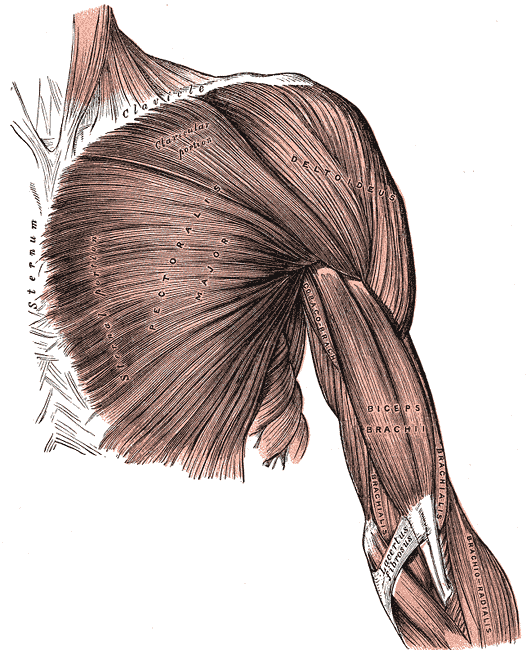
Pectoralis Major
The pectoralis major is the large, fan-shaped, most-superficial and most-powerful muscle of the pectoral region composed of a sternal and clavicular head. It forms part of the anterior axilla wall.
- Origin:
- Insertion: The distal attachment of both heads is into the lateral lip of the intertubercular sulcus of the humerus.
- Action:
- Adducts and internally rotates the upper limb and draws the scapula anteroinferiorly.
- The clavicular head also acts individually to flex the upper limb.
- Innervation: Lateral and medial pectoral nerves
- Lateral pectoral nerve arises from the lateral cord of the brachial plexus, cervical roots C5–C7
- Medial pectoral nerve arises from the medial cord of the brachial plexus, cervical roots C8, T1
- Arterial Supply: Pectoral branch of thoracoacromial (2nd part of axillary artery), perforating branches of internal thoracic artery (1st part of subclavian artery)
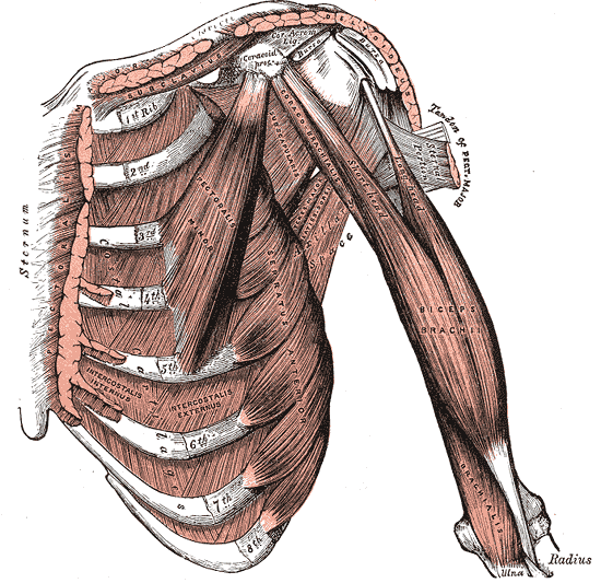
Pectoralis Minor
The pectoralis minor lies deep to the larger pectoralis major, and forms part of the anterior axilla wall.
- Origin: Originates from the 3rd-5th ribs
- Insertion: Inserts into the coracoid process of the scapula.
- Action: Stabilises the scapula by drawing it anteroinferiorly against the thoracic wall.
- Innervation: Medial pectoral nerve (arises from the medial cord of the brachial plexus, cervical roots C8, T1)
- Arterial Supply: Superior thoracic artery (1st part of axillary artery), thoracoacromial artery and lateral thoracic artery (both 2nd part of axillary artery)
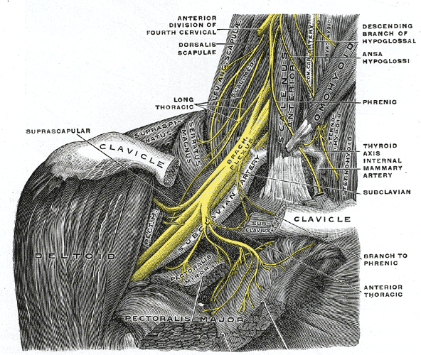
Serratus Anterior
The serratus anterior is located more laterally in the chest wall and forms the medial border of the axilla region.
- Origin: Consists of muscle slips originating from the lateral aspects of ribs 1-8
- Insertion: Costal surface of the medial border of the scapula
- Action: Rotates the scapula, allowing the arm to be raised over 90 degrees. It also holds the scapula against the ribcage
- Innervation: Long thoracic nerve (anterior rami of cervical roots C5, C6, C7)
Subclavius
The subclavius is a small triangular muscle between the clavicle and first rib. It forms part of the anterior wall of the axilla.
- Origin: First rib and cartilage
- Insertion: Subclavian groove of clavicle located on the inferior surface of middle third of the clavicle
- Action: Depression of the clavicle and elevation of the first rib
- Innervation: Subclavian nerve
- Arterial supply: Thoracoacromial trunk, clavicular branch (1st part of axillary artery)