

Overview
The adrenal (suprarenal) glands are paired endocrine glands situated over the medial aspect of the superior poles of each kidney in the retropertioneal space, between the superomedial kidney and the diaphragm. They secrete steroid and catecholamine hormones directly into the blood.
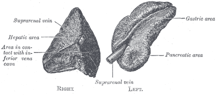
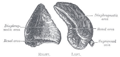
Structure
- Each gland has a body, two limbs (medial and lateral), outer connective tissue capsule and is enclosed in perirenal fascia, separated from the kidneys by perirenal fat.
- The perirenal fascia attaches the glands to the crura of the diaphragm.
- The right gland is pyramidal in shape, contrasting with the semi-lunar shape of the left gland.
- The adrenal cland consists an outer cortex and inner medulla
- Veins and lymphatics leave each gland via the hilum, but arteries and nerves enter the glands at numerous sites.
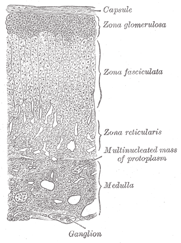
Cortex
The cortex derived from the embryonic mesoderm and is subdivided into three zones (from outer to inner):
- Zona glomerulosa – mainly produces and secretes mineralocorticoids such as aldosterone
- Zona fasciculata – mainly produces and secretes corticosteroids such as cortisol
- Zona reticularis – produces and secretes androgens such as dehydroepiandrosterone (DHES). It also secretes a small amount of corticosteroids.
Medulla
The medulla is derived from the ectodermal neural crest cells and consists of chromaffin cells which produces catecholamines (epinephrine, norepinephrine) as well as dopamine
Arterial Supply
The arterial supply is via three adrenal arteries:
- Superior adrenal artery – usually a group of 6-8 arteries arising from the inferior phrenic artery (from the abdominal aorta)
- Middle adrenal artery – arises from the abdominal aorta (often the largest of the adrenal arteries)
- Inferior adrenal artery – arises from the renal artery, a branch of the abdominal aorta at L1-L2
Venous Drainage
Adrenal veins emerge from the hilum and drain to different veins depending on the side:
- Left adrenal vein drains to the left renal vein or may join with the left inferior phrenic vein before entering the left renal vein
- Right adrenal gland drains to the IVC
Lymphatic Drainage
Lymphatics are contained within the capsule and drain to the lumbar and para-aortic nodes
Innervation
- Presynaptic sympathetic fibres – thoracic splanchnic nerves synapse directly with chromaffin cells
- Postsynaptic fibres – coeliac, aorticorenal, and renal ganglia innervate surrounding vessels
- Sympathetic innervation to the adrenal medulla is via myelinated pre-synaptic fibres, mainly from the T10 to L1 spinal cord segments.
Relations
Right Adrenal Gland
- Medial: Right crus of the diaphragm, right inferior phrenic artery
- Lateral: right lobe (bare area) of the liver
- Anterior: Inferior vena cava, parietal peritoneum
- Posterior: right kidne
Left Adrenal Gland
- Medial: Left crus of the diaphragm, left inferior phrenic artery
- Anterior: lesser sac, stomach, splenic artery, pancreas, parietal peritoneum
- Lateral: Spleen
- Posterior: Left kidney
Variants
- Lying down adrenal sign – takes on a flattened appearance
- Often in the presence of a pelvic kidney or renal agenesis
- Adrenal gland hypoplasia or agenesis
- Accessory adrenal rests often near adrenal glands but may be found anywhere in the abdomen, pelvis or scrotum
- Horseshoe adrenal gland