

Overview
The accessory nerve is the eleventh cranial nerve and has virtually only somatic motor function, innervating the sternocleidomastoid and trapezius muscles. It is thought to consist of a cranial (internal ramus) and spinal (external ramus) component and therefore has a double origin.

Origin
- Cranial component
- Derived from the nucleus ambiguus – a large motor neuron embedded in the reticular formation, which provides also motor fibres to the glossopharyngeal nerve and the vagus nerve.
- Arises from the ventrolateral surface of the medulla oblongat as 3 – 4 rootlets at the posterior border of the olive, inferior to the glossopharyngeal nerve and the vagus nerve.
- Smaller than the spinal component
- Spinal component
- Derived from the spinal accessory nucleus in the ventral horn of the upper five cervical segments.
- Rootlets arise from the lateral aspect of the upper five spinal cord segments and coalesce to form the solitary spinal component of the accessory nerve.
Course
Intracranial Segment
- Rootlets of the cranial component unify to form a single trunk, which runs laterally with glossopharyngeal nerve and vagus nerve to reach the jugular foramen where its joined by the spinal root of the accessory nerve at the lateral basal cistern.
- Rootlets of the spinal component unify and ascend in the vertebral canal to enter the posterior cranial fossa via the foramen magnum, traveling behind the vertebral artery and dorsal to the denticulate ligaments.
- Rootlets of the cranial and spinal components then connect to form the common trunk of the accessory nerve
- The common trunk turns upwards and travels laterally towards the jugular foramen
Skull Base Segment
- The accessory nerve passes through the posterior aspect of the pars vascularis portion of the jugular foramen.
- During passage through the jugular foramen, it runs through a dural sheath together with the vagus nerve laterally and the internal jugular vein anteriorly
- After the common trunk enters the retrostyloid space, it separates once again into fibres derived from cranial and spinal roots
- The cranial component forms connections with the superior ganglion of the vagus nerve at the level of the upper edge of the transverse process of C1, contributing to the vagal function of the pharyngeal and laryngeal area
- The spinal component innervates the trapezius and sternocleidomastoid muscles.
- As the accessory nerve exits the jugular foramen it is wrapped by the meningeal dura, which is attached to the periosteum.
Extracranial Segment
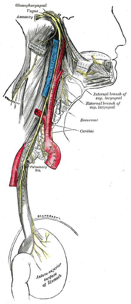
- The spinal component descends vertically within the carotid sheath between internal jugular vein and internal carotid artery
- The accessory nerve then travels past the transverse process of the atlas, anteriorly in most cases, though there is some variation; less commonly, it lies lateral or medial to the transverse process of the atlas
- At the level of carotid triangle, turns posteriorly, either superficial or deep to internal jugular vein, to pass deep to or through sternocleidomastoid, supplying it
- Enters posterior triangle, crossing it obliquely within investing layer
- Continues deep to surface of trapezius muscle, innervating it
Development
- Develops from the ganglionic crest (ectoderm) – the same origin as the vagus nerve
- At around 20 days, the ganglion crest is lateralised and divided into two portions (right and left)
- The caudal portion of the crest gives rise to a band of fibres at the level of the fourth, fifth and sixth cervical segment, which will become the future accessory nerve.
- During the fourth week, it frees itself from the link of the vagus nerve to throw itself into a mesodermal mass, which will be the sternocleidomastoid muscle.
- At the end of the fifth week of gestation, the nerve is located medial to the dorsal rootlets and is connected at irregular intervals to what will be the spinal cord. It connects with the first spinal ganglion.
- The cranial portion is a row of ganglia, found in the nerve once formed. The ganglia during development will be larger, forming a connection with the jugular ganglion of the vagus nerve.
- During the second and third months, the accessory nerve stretches and takes what will be its shape.
Variants
- Posterior root of C1 is absent and therefore does not anastomose with the anterior root of C1
- No connection between accessory nerve and posterior root of C1
Various anastomoses between accessory nerve and posterior roots of 1 - Accessory nerve crosses the internal jugular vein ventrally, dorsally, medially, laterally or passes through the vein
- Accessory nerve passes along the internal surface of the sternocleidomastoid and
- sends branches without penetrating the muscle (type A)
- a single branch, without penetrating the muscle (type B)
- penetrating the muscle deeply (type C).
- Trapezius muscle only innverated by the cervical roots.
- Intracranially, the accessory nerve anastomoses with the facial nerve to innervate both the sternocleidomastoid muscle.
- The cervical plexus can contribute to the function of the spinal accessory nerve, from the level of C2 and C3.
Clinical significance
- Can be injured during surgical intervention involving the posterior triangle, e.g. excisional biopsy of a cervical lymph node or central line insertion
References
https://anatomypubs.onlinelibrary.wiley.com/doi/full/10.1002/ar.23823