

Overview
The temporal bones are paired, dense, irregularly-shaped complex bones that comprise the lateral base of the skull. It contains the inner and middle ear, articulates with the temporomandibular joint of the jaw.
Structure
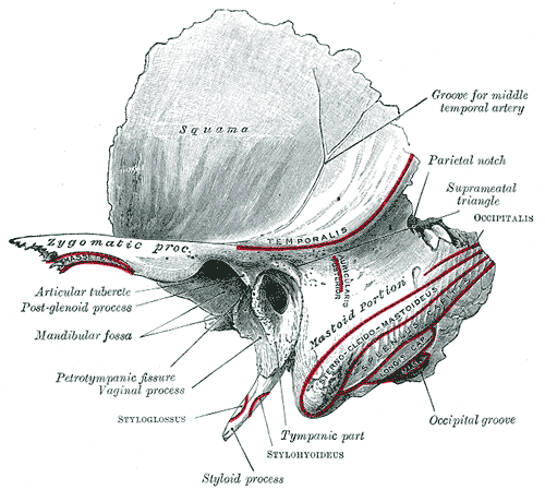
Squamous Part (squama temporalis)
- Largest part of the temporal bone
- Flat and plate-like, located anteriorly and superiorly
- The external surface is convex in shaping, forming part of the temporal fossa
- Grooved on the external surface for the middle temporal artery
- The internal surface forms the lateral part of the middle cranial fossa
- Has impressions that follow the groove and contour of the temporal lobe
- Grooved for the middle meningeal artery and vein
- The inferior aspect of the squamous part is the site of origin of the temporalis muscle
- Articulates with the sphenoid bone anteriorly and parietal bone laterally
- Zygomatic process – anterior projection from the squamous part of the temporal bone
Petrous Part
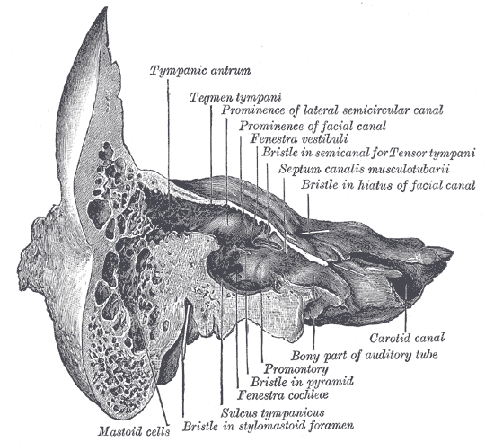
- Pyramidal-shaped bony mass located between the sphenoid and the occpital bones
- Forms the most medial part of the temporal bone
- Composes of a base, apex, anterior surface, posterior surface and inferior surface
- Contains the inner ear, acoustic labryinth
- The anterior region of the petrous part forms the posterior limit of the middle cranial fossa.
- Its internal surface is grooved by the inferior temporal gyrus and the trigeminal ganglion. The trigeminal impression is separated from another hollow posteriorly by a bony ridge. This hollow partially encloses the internal acoustic meatus and the cochlea.
- The ridge is limited posteriorly by the arcuate eminence, which is raised superiorly by the superior semicircular canal. The lateral aspect of the petrous part articulates with the squamous part of the temporal bone.
- Between this articulation laterally and the arcuate eminence medially, is a thin plate of bone called the tegmen tympani. This bone forms the roof of the middle ear. The lateral and posterior semicircular canals lie deep to the posterior slope of the arcuate eminence.
- The posterior area of the petrous part of the temporal bone contributes to the anterior limit of the posterior cranial fossa. It is continuous with the internal portion of the mastoid part. There is a depression below the arcuate eminence, the subarcuate fossa, which lies above the opening of the internal acoustic meatus.
- The inferior surface is highly irregular and contributes to the external surface of the cranial base.
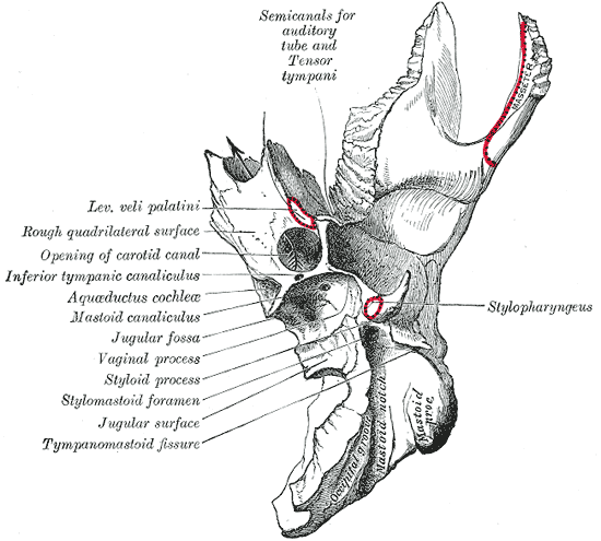
- Near the apex, there is a quadrangular area associated with the attachment of the levator veli palatini muscle and the pharyngotympanic tube. Posterior to this region is an opening for the carotid canal, and more posteriorly again is the jugular fossa.
- The superior border is the longest border of the petrous part of the temporal bone. It is inside the cranial cavity, and is grooved by the superior petrosal sinus.
- The posterior border forms part of the fossa for the inferior petrosal sinus. The anterior border is attached laterally to the squamous part of the temporal bone; medially it articulates with the greater wing of the sphenoid bone. Two canals exit the skull at the junction between the petrous and squamous parts: the upper one containing the tensor tympani muscle, the lower one containing the pharyngotympanic tube.
Mastoid Part
- Contains the mastoid air cells – acts as reservoir of air to equalise air pressure within the middle ear and lightens the weight of the calvarium
- Mastoid Process – inferior projection of bone which serves as the site of attachment for sternocleidomastoid
Tympanic Part
- Curved plate immediately below the origin of the zygomatic process
- Fuses with the petrous part internally, and the squamous and mastoid parts posteriorly.
- The anterior surface forms the posterior part of the mandibular fossa, and its lateral part forms part of the external acoustic meatus.
- Its concave posterior surface forms the anterior wall, floor and part of the posterior wall of the external acoustic meatus
- The central region is thin and often perforated
- The inferior border is sharp, and forms the vaginal process of the styloid process
- The stylomastoid foramen lies between the styloid process and the mastoid process. It is the terminal end of the facial canal, transmitting the facial nerve and the stylomastoid artery.
- Styloid process – a 2.5 cm narrow, usually-straight pointed projection that extends anteroinferiorly from the inferior surface of the temporal bone. The distal end is the site of a number of muscles and ligaments.
- The parotid gland lies lateral to the styloid process
- The facial nerve crosses its base;
- The external carotid artery passes through the parotid gland crossing the tip of the styloid process
- Medially is the attachment of the stylopharyngeus muscle, separating the styloid process from the internal jugular vein.
Mastoid Part
- Most posterior part
- Its outer surface is roughened by muscular attachments.
- There is a downward conical projection called the mastoid process from the mastoid part.
- A number of muscles are attached to the mastoid process, these being the sternocleidomastoid, splenius capitis and longissimus capitis muscles.
- Mastoid notch – a depression on the medial surface of the mastoid process onto which the posterior belly of the digastric is inserted.
- The medial surface of the mastoid process has a deep groove called the sigmoid sulcus, in which the sigmoid sinus is located.
Articulations
- Temporomandibular joint
- The squamous part of the temporal bone articulates with the sphenoid bone anteriorly and the parietal bone laterally.
- The zygomatic process of the temporal bone also articulates with the zygomatic bone to form the zygomatic arch (i.e. cheekbones).
Foramina
- Foramen lacerum – great petrosal nerve
- Carotid canal – interal carotid artery
- Internal acoustic meatus – facial nerve (CN VII), vestibulocochlear (CN VIII), labyrinthine artery
- Jugular foramen – internal jugular vein, glossopharyngeal nerve (CN IX), vagus nerve (CN X), accessory nerve (CN XI), posterior meningeal artery
Muscular Attachments
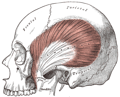
- Description: fan-shaped muscle covering temporal, frontal and parietal bones
- Origin: temporal fossa
- Insertion: coronoid process of mandible
- Action: closes jaw, elevates and retracts mandible
- Nerve supply: trigeminal nerve
- Description: powerful muscle covering lateral aspect of mandibular ramus, composed of superficial and deep head
- Origin: zygomatic arch and maxilla
- Insertion: angle and ramus of mandible
- Action: primer mover of jaw closure; elevates mandible
- Relations:
- Lateral to mandible
- Nerve supply: trigeminal nerve
Sternocleidomastoid
- Description: two-headed neck muscle
- Origin: Manubrium and medial portion of the clavicle
- Insertion: Mastoid process of the temporal bone, superior nuchal line
- Action:
- Unilaterally: contralateral cervical rotation, ipsilateral cervical flexion
- Bilaterally: cervical flexion, elevation of sternum and assists in forced inhalation.
- Relations:
- Lateral to mandible
- Nerve supply:
- Motor: spinal accessory nerve
- Sensory: cervical plexus
- Proprioceptive: ventral rami of C2-3
The digastric consists of two muscular bellies connected by an intermediate tendon:
- Attachments:
- Anterior belly arises from the digastric fossa of the mandible
- Posterior belly arises from the mastoid process of the temporal bone
- The two bellies are connected by an intermediate tendon, which is attached to the hyoid bone via a fibrous sling
- Actions: Depresses the mandible and elevates the hyoid bone.
- Innervation:
- Anterior belly – inferior alveolar nerve, a branch of the mandibular division of the trigeminal nerve
- Posterior belly – digastric branch of the facial nerve
Splenius Capitis
- Description: a broad, straplike muscles in the back of the neck
- Origin: lower half of the nuchal ligament, from the spinous process of the seventh cervical vertebra, and upper three-four thoracic vertebrae
- Insertion: mastoid process of the temporal bone, deep to the sternocleidomastoid muscle
- Action: head extension, also lateral flexion and rotation of the cervical spine (shaking the head)
- Relations:
- Deep to sternocleidomastoideus at the mastoid process and to the trapezius for its lower portion.
- Forms the floor of the posterior triangle of the neck
- Nerve supply: posterior ramus of the spinal nerves C3 and C4