

Overview
A horseshoe-shaped bone in the anterior midline of the neck at the C3 vertebral level (the same level of the lateral glossoepiglottic folds within the pharynx and defines the junction of the oropharynx and larygngopharynx). It serves as a strut or anchor for attachment of muscles of the floor of the mouth superiorly (e.g. tongue) and anterior neck inferiorly, such that it is suspended and has no bony articulations, to aid predominantly in tongue movement and swallowing.
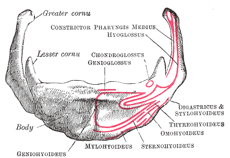
Structure
- Body – central portion, has an anterior convex surface
- Greater horn (cornu) – projects superolaterally from each end of the body
- Lesser horn (cornu)- smaller conical eminences, projects superoposteriorly from the superior aspect of the body

[foogallery id=”7850″]
Muscular Attachments
Superior
- Middle pharyngeal constrictor muscle – attaches to the length between the lesser and greater horns

Extrinsinc tongue muscles
- Hyoglossus muscle
- Origin: Body and greater horn of hyoid bone
- Insertion: Lateral and inferior aspect of tongue
- Innervation: Terminal lingual nerves of the hypoglossus nerve
- Genioglossus
- Origin: Mental spine of mandible
- Insertion: Dorsum of tongue, hyoid bone
- Innervation: Terminal lingual nerves of the hypoglossus nerve
Suprahyoid muscles
- Digastric muscle
- Origin: Anterior belly – digastric fossa of mandible, Posterior belly – mastoid notch of temporal bone
- Insertion: Intermediate tendon attached to body of hyoid
- Stylohyoid muscle
- Origin: Posterior border of styloid process
- Insertion: Body of hyoid bone at junction with greater horn
- Geniohyoid muscle
- Origin: Inferior genial tubercle on back of symphysis mentis of the mandible
- Insertion: Anterior surface of body of hyoid bone
- Innervation: Hypoglossus nerve
- Mylohyoid muscle
- Origin: Mylohyoid line of mandible
- Insertion: Median raphe and body of hyoid bone
Infrahyoid muscles
- Thyrohyoid muscle (deepest)
- Origin: Oblique line on lamina of thyroid cartilage
- Insertion: Lower border of body and greater horn of hyoid bone
- Innervation: Branch from anterior rami of C1 spinal nerve
- Omohyoid muscle (anterior, lateral)
- Origin: Inferior belly – from upper border of scapula and superior transverse scapular ligament, ending in intermediate tendon, Superior belly – intermediate tendon
- Insertion: Inferior belly – intermediate tendon, Superior belly – body of hyoid bone
- Innervation: Superior root of the ansa cervicalis formed by hypoglossus nerve
- Sternohyoid muscle (anterior, medial)
- Origin: Posterior surface of manubrium of the sternum, posterior sternoclavicular ligament, medial
end of clavicle - Insertion: Medial part of lower border of body of hyoid bone
- Innervation: Superior root of the ansa cervicalis formed by hypoglossus nerve
- Origin: Posterior surface of manubrium of the sternum, posterior sternoclavicular ligament, medial
Ligamentous Attachments
- Stylohyoid ligament – extends from the styloid process of the temporal bone to the lesser horns, lateral to the posterior belly of diagastric muscle and anterior to the posterior belly.
- Thyrohyoid membrane – extends from the superior border of the thyroid cartilage to the posterior surface of hyoid and lower border of greater horns. It is pierces by the interna laryngeal nerve and superior laryngeal artery (branch of superior thyroid artery).
- Median thyrohyoid ligament – the thickened medial part
- Hyoepiglottic ligament – extends from the anterior aspect of the epiglottis to the rounded upper body of the hyoid bone and raises the oral mucous membrane to form the median glossoepiglottic fold. Fibrous tissue also passes from the greater horn to the side of the epiglottis and raises the lateral glossoepiglottic fold.
Blood supply
- Supplied via the lingual artery, which courses from the tongue to the greater horns
- The suprahyoid branch of the lingual artery runs along the upper border of the hyoid bone and supplies blood to the attached muscles
Relations
- Posteriorly
- Between the posterior surface of the body and the thyrohyoid membrane is a bursa
- Upturned loop of the thyroglossal duct
- Epiglottis
- Anteriorly
- Separates cervial lymph node levels Ia (above) from VI (below)
- Layers (superficial to deep): Skin, superficial fascia, platysma, superficial layer of deep cervical fascia
- Anterior jugular vein – descends on the surface of sternohyoid muscle, drains into external jugular vein via the jugular venous arch
- Thyroglossal duct
- Laterally
- Separates cervial lymph node level II from level III
- Lateral to the greater horns: external carotid artery, ansa cervicalis, internal jugular vein
- Superiorly
- Superior to the greater horns: submandibular gland
- Mandible
- Inferiorly
- Pretracheal layer – encapsulates the thyroid gland
- Thyroid cartilage
- Superior laryngeal artery (branch of superior thyroid artery arising from external carotid artery, pierces the thyrohyoid membrane to supply intrinsic muscles of the larynx)
- Superior laryngeal nerve – branch of the inferior ganglion of the vagus nerve. Divides into internal and external branches . Internal laryngeal nerve – enters the larynx through the thyrohoid membrane and supplies mucosa above the glottis, External laryngeal nerve – travels to the inferior pharyngeal constrictor muscle. Innervates cricothyroid muscle
Anatomical Variants
- Various degrees of ankylosis
- Hyoid bone body exostoses
- Unilateral/bilateral hypo/hyperplasia of lesser or greater horn
- Accessory bones
- Unilateral/bilateral absence of lesser or greater horn
- Triticeal cartilage
- Non-fusion of the superior horn of the thyroid to the thyroid cartilage (can mimic a fracture)
- Articulating connection between greater horn and superior horn of the thyroid cartilage.
- Nearly circumferential ankylosed hyoid bone (rare)
- Elongation of the styloid process; Eagle’s syndrome.
Development
- The second pharyngeal arch gives rises to the lesser horn and upper part of the body
- The cartilage of the third pharyngeal arch forms the greater cornu and lower portion of the body
Imaging
- Fluoroscopic swallow study – the hyoid bone moves superiorly and anteriorly during a swallow. The degree of movement indirectly indicates the strength of pharyngeal musculature, where lack of movement suggests pharyngeal paresis.
References
Variants of the hyoid-larynx complex, with implications for forensic science and consequence for the diagnosis of Eagle’s syndrome
https://www.nature.com/articles/s41598-019-52476-z#:~:text=Anatomical%20variations%20of%20the%20hyoid%2Dlarynx%20complex%20First%20vertical%20column,%3A%20ventral%2C%20D%3A%20dorsal.
https://www.nature.com/articles/s41598-019-52476-z#:~:text=Anatomical%20variations%20of%20the%20hyoid%2Dlarynx%20complex%20First%20vertical%20column,%3A%20ventral%2C%20D%3A%20dorsal.