

Overview
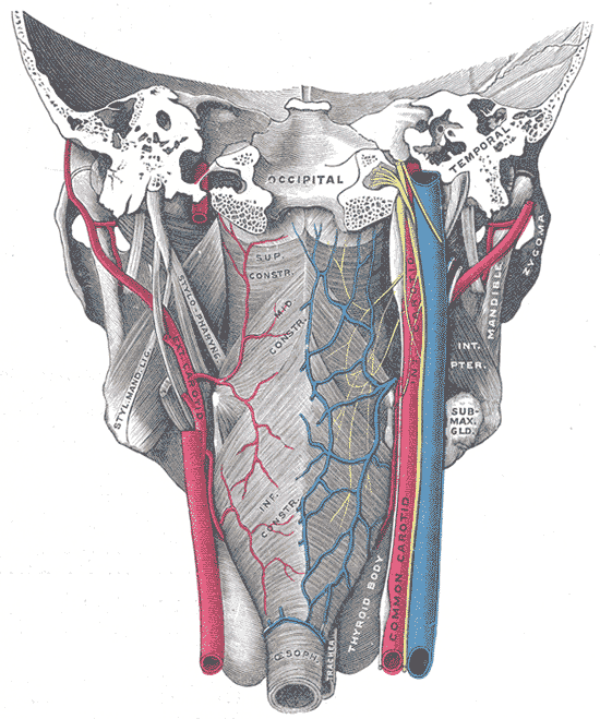
The pharynx is a muscular tube that connects the nasal and oral cavity to the larynx and oesophagus. Three overlapping pairs of muscles form the pharyngeal wall, extending from the skull base to the the inferior border of the cricoid cartilage (anteriorly) and C6 (posteriorly).
The pharynx is comprised of three parts (superior to inferior):
- Nasopharynx
- Oropharynx
- Laryngopharynx

Structure
The pharyngeal muscles (involuntary skeletal) are organised into two muscular layers – the outer circular layer and the inner longitudinal layer. They are all innervated by the pharyngeal plexus supplied by the vagus nerve with the exception of the stylopharyngeus muscle which is innervated by the glossopharyngeal nerve.
Outer circular layer
During swallowing, these muscles constrict to propel bolus downwards (an involuntary process termed peristalsis). It includes:
- Superior constrictor muscle
- Origins: Pterygoid hamulus, pterygomandibular raphe, posterior end of mylohyoid line of mandible
- Insertions: Pharyngeal tubercle on basilar part of occipital bone
- Middle constrictor muscle
- Origins: Stylohyoid ligament, greater and lesser horns of the hyoid bone
- Insertions: Median pharyngeal raphe, blends with superior and inferior pharyngeal constrictors
- Inferior constrictor muscle
- Origins: Thyropharyngeal part: Oblique line of thyroid cartilage, Cricopharyngeal part: Cricoid cartilage
- Insertions: Thyropharyngeal part: Median pharyngeal raphe, Cricopharyngeal part: Blends inferiorly with circular esophageal fibres
Inner longitudinal layer
During swallowing, these muscles act to shorten and widen the pharynx. It includes:
- Stylopharyngeus muscle – innervated by glossopharyngeal nerve
- Origin: Medial base of styloid process of temporal bone
- Insertion: Blends with pharyngeal constrictors, lateral glossoepiglottic fold, posterior border of thyroid cartilage
- Salpingopharyngeus muscle
- Origin: Inferior/cartilaginous part of auditory (Eustachian) tube
- Insertion: Blends with palatopharyngeus muscle
- Palatopharyngeus muscle
- Origins: Posterior border of hard palate, palatine aponeurosis
- Insertions: Posterior border of thyroid cartilage, blends with contralateral palatopharyngeus muscle
Supportive Structures
- Pharyngobasilar fascia – strong internal fascial lining. Superior constrictor does not extend up to skull base. This fascia bridges the gap and forms the posterior wall of nasopharynx.
- Attaches to basilar occipital and petrous temporal bone
- Pharyngeal Tubercle – Tubercle on inferior surface of basiocciput, anterior to foramen magnum.
- Pharyngeal raphe – midline raphe extending from pharyngeal tubercle to oesophagus at C6.
Arterial Supply
Predominantly supplied by facial artery, which gives the tonsillar artery and supplies the tonsils. Other branches include:
- The ascending pharyngeal artery
- The lingual artery
- The ascending and descending palatine arteries
Venous Drainage
- External palatine vein – drains into the pharyngeal venous plexus and from there into the internal jugular vein.
Lymphatic Drainage
- Jugulodigastric node
- Superior and inferior deep cervical nodes
Anatomical Variants
- Pharyngeal diverticuli