

RANZCR Syllabus
Category 1
• External auditory meatus
• Tympanic membrane
• Mastoid air cells
Category 2
Nil
Category 3
• Auricle & its innervation
• Tympanic ring
• External auditory meatus
• Tympanic membrane
• Mastoid air cells
Category 2
Nil
Category 3
• Auricle & its innervation
• Tympanic ring
External auditory meatus |
|---|
| CATEGORY 1NOT EXAMINED |
A sigmoid-shaped oval-cylindrical canal canal extending from bottom of the concha to the tympanic membrane.
Course
- Pars externa – Directed inward, forward and slightly upward
- Pars media – Passes inward and backward
- Pars interna – Passed inward, forward and slightly downward
Relations
- Anteriorly: condyle of the mandible
- Posteriorly: mastoid air cells
Blood supply
- Branches from the posterior auricular, internal maxillary and temporal
Innervation
- Auriculotemporal branch of the mandibular nerve and auricular branch of vagus.
Tympanic Membrane |
|---|
| CATEGORY 1MAR 2015 |
A thin, 1cm diameter cone-shaped membrane separating the external ear from the middle ear. Its primary function is to transmit sound from the air to the ossicles of the middle ear.
Orientation
- Oriented obliquely at 55 degrees with the external acoustic meatus
- Faces downwards, forwards and laterally.
Relations
- Superiorly: middle cranial fossa
- Inferiorly: parotid gland
- Anteriorly: temporomandibular joint
- Posteriorly: ossicles and facial nerve.
Regions
- Pars flaccida – small, fragile portioning. Located above the lateral process of the malleus
- Pars tensa – three layers (skin, fibrous tissue and mucosa). Thickened edge forms a fibrocartilaginous ring called the annulus tympanicus (Gerlach’s ligament). The central umbo tents inward towards the level of the tip of the malleus. The middle fibrous layer encloses the handle of the malleus.
Blood supply
- Internal (mucosal) surface – stylomastoid artery (posterior auricular) forms a circular anastomosis with the anterior tympanic branch of maxillary round the margin of the membrane
- External (meatal) surface – deep auricular artery (maxillary).
Innervation
- Internal (mucosal) surface – tympanic branch of the glossopharyngeal nerve via the tympanic plexus
- External (meatal) surface – auriculotemporal nerve (branch of CN VIII), supplemented by auricular branch of the vagus.
Mastoid Air Cells |
|---|
| CATEGORY 1MAR 2015 |
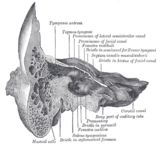
A collection of air-filled spaces in the mastoid process of the petrous portion of the temporal bones. The air cells help to:
 function as sound receptors
- provide voice resonance
- act as acoustic insulation and dissipation
- provide protection from physical damage
- reduce the mass of the cranium.
Structure
The air cells are contained within the mastoid antrum, a cavity communicating posteriorly with the middle ear and anteriorly with the epitympanic recess of the middle ear.
- Superiorly: air cells are large and irregular
- Inferiorly: air cells diminish in size
- Apex: small and containing marrow, otherwise entirely absent (i.e. solid).