

• Lacrimal sac & duct
Category 2
• Lids & tarsal plates
• Blood supply & venous drainage
• Levator palpebrae superioris & nerve supply
• Conjunctival sac boundaries
• Lacrimal canaliculi
Category 3
• Lateral and medial check ligaments of the globe
Overview
Tears produced by the lacrimal gland, accessory lacrimal glands of Krause, Wolfring and Zeis, and Meibomian glands track medially along the eyelid margins and collect at the lacrimal lake at the inner canthus. The nasolacrimal drainage apparatus drains tears in a descending course through the inferior and superior lacrimal canaliculi and common canaliculus through the valve of Rosenmuller leading to the lacrimal sac, which drains into the nasolacrimal duct in the nasolacrimal canal of the maxilla and through the valve of Hasner to the inferior nasal meatus below the inferior turbinate in the nasal cavity
The upper dilated end of the nasolacrimal duct, located in a groove formed by the lacrimal bone and frontal process of the maxilla. It functions as a reservoir for tears.
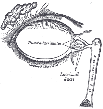
Structure
- Generally ovoid in shape measuring 12 to 15mm
- Superior end: closed and round
- Inferior end continuous with the nasolacrimal duct
Lacrimal Canaliculi
- Superior and inferior and commence at the respective lacrimal puncta
- Drains tears from lacrimal lake at the inner canthus (medial angle) of the eye.
- Unite to form common canaliculus and drain via sinus of Maier into lacrimal sac, between medial palpebral ligament and orbital septum.
Lacrimal Sac
- Lies in lacrimal fossa, between anterior and posterior lacrimal crests.
- Drains via valve of Krause into nasolacrimal duct.
Nasolacrimal Duct
- Slopes inferiorly into the inferior meatus of nasal cavity
- Mucous membrane raised into several folds to form Valve of Hasner.
- Two parts – intraosseous within nasolacrimal canal of maxilla, and membranous part which runs in nasal mucosa.