

Overview
The spinal cord is a tubular bundle of nervous tissue (grey matter internally and white matter externally) and supporting cells, located within the vertebral canal of the vertebral column.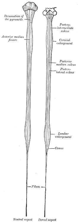
Internal structure
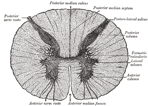
A transverse section of the spinal cord demonstrates a peripheral mass of white matter surrounding a butterfly-shaped central part of the spinal cord composed of gray matter (containing neuronal cell bodies). Centrally, there is a small ependyma-lined longitudinal central canal filled with CSF which is continuous with the fourth ventricle caudally.
Grey Matter
The grey matter contains the cell bodies of neurons and glia.
- Anterior horns – contain motor neurons for skeletal muscles, sends efferent fibre via the ventral nerve roots
- Lateral horns – contains autonomic neurons for pelvic and visceral organs, only present in the thoracic region
- Dorsal horns – contains somatosensory neurons, receives primary afferents from the dorsal roots of spinal nerves
- Periependymal grey matter – divided into ventral and dorsal grey matter commissures
White Matter
The white matter contains nerve fibres of tracts and is divided into anterior, dorsal and lateral columns (also known as funiculi), as well as the anteior spinal commisure.
- Anterior columns – contain the spinothalamic tracts (responsible for pain, temperature, non-discriminative touch and pressure sensation)
- Dorsal columns – contain ascending fibres (responsible for vibration, conscious propioception and discriminative touch sensation)
- Lateral columns – contain the corticospinal tracts (principal motor pathway connecting the cerebral cortex to spinal motor neurons)
- Anterior spinal commissure – located between the posterior-most extent of the anterior median fissure anteriorly and the ventral grey matter commissure posteriorly
External Structure

- The cord is sheathed in the same three meninges as is the brain: the pia, arachnoid and dura. The dura is the tough outer sheath, the arachnoid lies beneath it, and the pia closely adheres to the surface of the cord
- The spinal cord is attached to the dura by a series of lateral denticulate ligaments emanating from the pial folds
- Anterior median fissure – deep groove extending the length of the anterior surface of the spinal cord.
- Posterior median fissure –
- Spinal nerves – mixed nerves that originate from the spinal cord, forming the peripheral nervous system.
- Anterior and posterior roots arise from the spinal cord within the vertebral canal, and unite at the intervertebral foramina, forming a single spinal nerve, then divides into two rami:
- Anterior rami – supplies nerve fibres to much of the remaining area of the body, both motor and sensory.
The nerve roots L2-S5 arise from the distal end of the spinal cord, forming a bundle of nerves known as the cauda equina. - Posterior rami – supplies nerve fibres to the synovial joints of the vertebral column, deep muscles of the back, and the overlying skin.
- Anterior rami – supplies nerve fibres to much of the remaining area of the body, both motor and sensory.
- Anterior and posterior roots arise from the spinal cord within the vertebral canal, and unite at the intervertebral foramina, forming a single spinal nerve, then divides into two rami:
Course
- Arises cranially at the foramen magnum at the base of the skull as a continuation of the medulla oblongata
- Descends within the vertebral canal, surrounded by spinal meninges
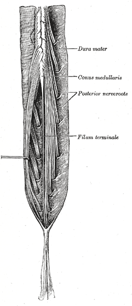
- At the level of L1-L2, the spinal cord tapers forming the conus medullaris
Termination
- A thin thread filum terminale extends from the tip of the conus medullaris to the 1st coccygeal vertebra, anchoring the spinal cord in place
Segments
The cord is organised into 31 segments, defined by the 31 pairs of nerves exiting the cord
Cervical
- 8 cervical nerve roots
- Cervical enlargement
- Symmetric enlargement between segmental levels C5 to T1, with the greatest circumference occurring at C6
- The source of the spinal nerves that contribute to the brachial plexus and supply the upper limbs, thus contains increased volume of motor cells in the ventral horns of the grey matter
Thoracic
- 12 thoracic nerve roots
Lumbar
- 5 lumbar nerve roots
- Lumbar enlargement
- Symmetric enlargement between segmental levels T11 to the conus medullaris
- The source of the spinal nerves which contribute to the lumbar and sacral plexuses
- Cauda equina (Latin: horse’s tail)
- Bundle of spinal nerves and spinal nerve rootlets contained within the lumbar cistern (subarachnoid space), which result from the termination of the spinal cord as conus medullaris at the level of L1-L2
- Nerve roots of spinal nerves of L2 to S5 (including the coccygeal nerve) descend to exit via neural foramina at their respective vertebral levels, arranged such that lumbar roots are more lateral and the coccygeal roots are medial
Sacral
- 5 sacral and 1 coccyx nerve roots
Arterial Supply
The spinal cord is supplied by extensive collateral supply formed by branches of the vertebral and segmental arteries. It is predominantly supplied by three longitudinal arteries (single anterior spinal artery and paired posterior spinal arteries):
- Anterior spinal artery
- Origin: Formed by branches of the vertebral artery before they unite to form the basilar artery.
- Course: Anterior spinal artery arises from two small branches, arising bilaterally near the termination of the vertebral artery. The branches descends along the anterior aspect of the medulla oblongata, uniting at the foramen magnum to form a single trunk. The anterior spinal artery travels in the anterior median fissure, terminating as a slender twig on the filum
- Supplies: Anterior two-thirds of the spinal cord.
- Posterior spinal artery (paired)
- Origin: Arise mostly from PICA (sometimes vertebral artery).
- Course: Descends posterior to the medulla, passing in front of the posterior roots of the spinal nerves.
- Supplies: Posterior one-third of the spinal cord
Reinforced by radicular or segmental medullary arteries which enter the vertebral canal via the intervertebral foramina and divide into anterior and posterior branches with variable anastomoses with the spinal arteries. The anterior and posterior spinal arteries are connected by pial anastomoses termed arterial vasocorona. The radicular and segmental arteries include:
- PICA, vertebral arteries, ascending cervical arteries, deep cervical arteries, posterior intercostal arteries, lumbar arteries, median and lateral sacral arteries.
- Artery of Adamkiewicz – The dominant segmental artery of the lumbosacral cord segments is the artery of Adamkiewicz, arising on the left sides between T9 and L2.
Venous Drainage
- Drain into segmental radicular and medullary veins which connect segmentally via intervertebral veins into systemic veins and internal and external vertebral plexuses
- Drain into lumbar and sacral veins which drain into median sacral vein, internal and common iliac veins and inferior vena cava
Development
- Stops growing at age of 4, whilst the vertebral column finishes growing at age 14 – 18
Anatomical Variants
- Variable origin of artery of Adamkiewicz
- arises on the right side
- arising from lumbar arteries at level of L1 – L2, intercostal arteries T5 – T8
- Duplicate anterior spinal arteries
- Single-origin anterior spinal artery from unilateral vertebral artery
- Single or duplicate anterior spinal arteries arising off vascular arcade between vertebral arteries
- Posterior spinal arteries arising from vertebral artery (25%) or PICA (75%)