

Overview
The atlas (C1) is the first cervical vertebra, consider an atypical vertebra.

Structure
- Consists of paired lateral masses joined by anterior and posterior arch
- No vertebral body, fuses to C2 during development to become dens
- Lateral mass – paired, ovoid shaped featuring:
- Superior articular surface – bean shaped and concave, articulates with occipital
condyle (atlanto-occipital joint) allowing nodding - Inferior articular process – circular and flat, articulates with superior facet of C2
(lateral atlanto-axial joint) - Medial surface – marked by vascular foramin and tubercle for attachment of the transverse ligament
- Superior articular surface – bean shaped and concave, articulates with occipital
- Anterior arch
- Facet on posterior surface for articulation with dens (median atlanto-axial joint)
- Anterior tubercle – attachment of anterior longitudinal ligament and anterior atlanto-occipital membrane
- Posterior arch
- Longer than anterior arch, constituting 3/5 of the circumference of the ring
- Posterior tubercle for attachment of ligamentum nuchae
- Upper surface grooved by vertebral artery posterior to facet for occipital condyle
- Superior border for attachment of posterior atlanto-occipital membrane
- Inferior border for attachment of ligamentum flava
- Transverse process – large, protrude further laterally than other cervical vertebra
- Foramen transversarium contains vertebral artery, vertebral vein and sympathetics
Articulations
- Atlanto-occipital joint: hyaline-covered synovial joint between the occipital condyle and concave facet of the lateral mass of the atlas. Covered by a capsule and innervated by C1, this joint allows for flexion, extension and lateral flexion.
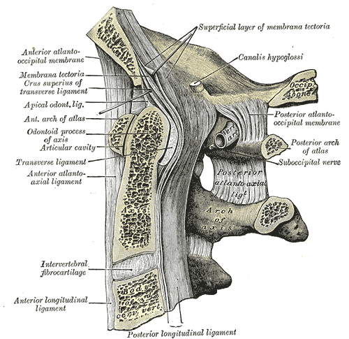
- Median atlantoaxial joint: hyaline-covered synovial joint between the dens of the axis and the posterior aspect of the anterior arch of the atlas, allowing for the rotation of the head. The dens is held in place by the transverse ligament, with a bursa between the two.
- Lateral atlantoaxial joint: hyaline-covered synovial joint between the inferior articular facet of the atlas and the superior articular facet of the axis which allows for the rotation of the head. A capsule innervated by the C2 nerve surrounds the joint.
Ligamentous Attachments
- Transverse ligament – Strong band that runs posterior to the dens of the axis, holding it in place. Each end is attached to tubercles on the anterior arch of the atlas. Forms part of the cruciate ligament, along with the superior and inferior longitudinal ligaments.
- Atlantoaxial ligaments: attach from the lower border of the anterior arch of the atlas to front of the body of the axis. Provides tertiary support against ventral translation of the dens.
Tendinous Attachments
- Anterior atlanto-occipital membrane: attached to upper border of the anterior arch to the outer margins of foramen magnum
- Posterior atlanto-occipital membrane: attached to upper border of the posterior arch to the outer margins of foramen magnum. At each lateral margin, there is a gap for the passage of the C1 nerve and vertebral artery, which sometimes ossifies and becomes a foramen. Innervated by C1.
Muscular Attachments
- Longus colli – Anterior tubercle
- Levator scapulae – Tip of the transverse processes
- Splenius cervicis – Transverse processes
- Obliquus capitis superior – Transverse processes. Forms superolateral border of the suboccipital triangle.
- Obliquus capitis inferior – Transverse processes. Forms inferolateral border of the suboccipital triangle.
- Rectus capitis posterior minor – Tubercle on the posterior arch of the atlas
Development
- The atlas ossifies from three centers
- Of these, one appears in each lateral mass about the seventh week of fetal life, and extends backward; at birth, these portions of bone are separated from one another behind by a narrow interval filled with cartilage.
- Between the third and fourth years they unite either directly or through the medium of a separate center developed in the cartilage.
- At birth, the anterior arch consists of cartilage; in this a separate center appears about the end of the first year after birth, and joins the lateral masses from the sixth to the eighth year.
- The lines of union extend across the anterior portions of the superior articular facets.
- Occasionally there is no separate center, the anterior arch being formed by the forward extension and ultimate junction of the two lateral masses; sometimes this arch is ossified from two centers, one on either side of the middle line
Anatomical Variants
- Arcuate foramen: calcification of the posterior atlanto-occipital membrane
- Superior articular facets of the lateral masses can be divided into two parts, with the anterior part being larger and the posterior smaller
- Fusion defects – incomplete anterior arch or posterior arch fusion
- Central or paramedian parts of the posterior arch can be absent and replaced by fibrous tissue
- Split atlas – rare congenital fusion defects of both the anterior and posterior arches
- Fusion (with the occiput – partially or fully)
- Epitransverse process of the atlas
- Clefts in arch
- Aplasia or hypoplasia