

Overview
Lymphatic drainage of breast is the primary route for metastatic dissemination of adenocarcinoma. The breast lymphatics are diffuse and variable, but generally it originates from breast lobules and flows through intramammary nodes and channels into a subareolar plexus, called Sappey’s plexus. From this plexus, lymphatic drainage takes place through three main routes that parallel venous tributaries. Lymphatics from the left breast ultimately terminate in the thoracic duct and the left subclavian vein, and from the right breast in the right subclavian vein.
Classical categorisation
There are four classically described intercommunicating lymphatic plexi of the breast, categorised as superficial or deep plexi.
Superficial Plexi
- The two superficial plexi are located in the dermis (cutaneous plexus) and in the superficial subcutaneous region (subcutaneous plexus).
- Superficial plexi may drain directly to the axillary lymph nodes
- Obstruction of the dermal channels by emboli of carcinoma cells causes oedema of the skin known as peau d’ orange – characteristic of inflammatory breast carcinoma
Deep Plexis
- One of the deep plexi is located in the pectoralis major muscle fascia (fascial plexus)
- The other deep plexus is located in the mammary gland, lobes and ducts (glandular plexus)
- Deep plexi may drain directly to axilla or alternatively:
- Initially drain into intramammary and interpectoral lymph nodes
- Drain into parasternal (“internal mammary”) lymph nodes
The glandular plexus communicates via lymphatic vessels that accompany the lactiferous ducts with a subareolar plexus known as Sappey’s plexus. Early research postulated that all breast lymph drained initially to the subareolar plexus, however, subsequent studies have demonstrated alternative routes.

Axillary (lateral) pathway
- The majority of the lymphatic drainage (>75%) of the breast is to the ipsilateral axillary nodes.
- Dominant pathway (receives >75% of lymph from breasts)
- Drains lateral quadrants of breast either directly or via Sappey’s plexus to axillary nodes
- Either runs around inferior border of pectoralis major to reach the pectoral group of lymph nodes or pass directly to the subscapular group
- Few channels from superior breast to the apical group sometimes interrupted by the infraclavicular group of lymph nodes
Internal mammary pathway
- Originates from both the medial and lateral quadrants of the breast
- Passes through the intercostal spaces and pectoralis major into parasternal/internal mammary lymph nodes
- Connections may lead across the median plane and hence to the contralateral breast
Retromammary pathway
- Comes from the deeper portion of the breast
- Drains to the subclavicular plexus
Other pathways occur when usual channels are blocked in disease. Lymph may pass to the contralateral breast, cervical nodes, peritoneal cavity and liver through the diaphragm or through the rectus sheath.
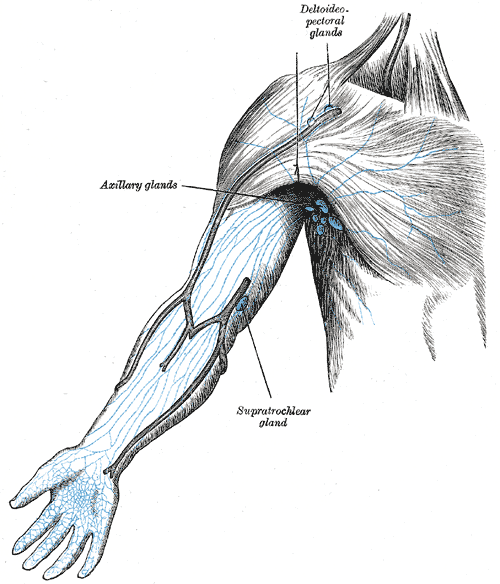
Axillary Groups
There are five axillary lymph node groups, namely the lateral (humeral), anterior (pectoral), posterior (subscapular), central and apical nodes. The apical nodes are the final common pathway for all of the axillary lymph nodes.
Lateral (humeral) lymph nodes
- Location: Posteromedial to axillary vein
- Receives: Upper limb
- Drains into: Central, apical and deep cervical nodes
Anterior (pectoral) lymph nodes
- Location: Inferior border of pectoralis minor, near lateral thoracic vessels
- Receives: Skin and muscles of the supraumbilical anterolateral body wall, breast
- Drains into: Central and apical nodes
Posterior (subscapular) lymph nodes
- Location: Along subscapular vessels on inferior margin of the posterior axillary wall
- Receives: Skin and muscles of the posterior and inferior body wall
- Drains into: Central and apical nodes
Central lymph nodes
- Location: Fat of the axilla
- Receives: The above mentioned lateral, anterior and posterior lymph node groups
- Drains into: Apical nodes
Apical (terminal) lymph nodes
- Location: Posterior and superior to pectoralis minor, towards the axillary vein
- Receives: Cephalic vein nodes, upper peripheral breast, the above mentioned central nodes
- Drains into: Subclavian trunk (may drain into jugulosubclavian venous trunk, subclavian vein, jugular lymphatic trunk, right lymphatic duct (left into thoracic duct), inferior deep cervical nodes)
Surgical Levels
The axillary lymph nodes drain about 75% of breast tissue and are divided into 3 surgical levels based on their relationship with the pectoralis major muscles. A traditional axillary lymph node dissection usually removes nodes in levels I and II, usually with a mastectomy if the breast cancer invasive.
Level 1
- Bottom level – lying below the inferior margin of the pectoralis minor
- Lymph nodes located below lateral to the lateral margin of the minor pectoralis muscle
Level 2
- Lymph nodes located behind the pectoralis minor (i.e. between the medial and lateral margins) and the interpectoral lymph nodes
Level 3
- Lymph nodes located between the upper/medial border of pectoralis minor and lower border of the clavicle