

Overview
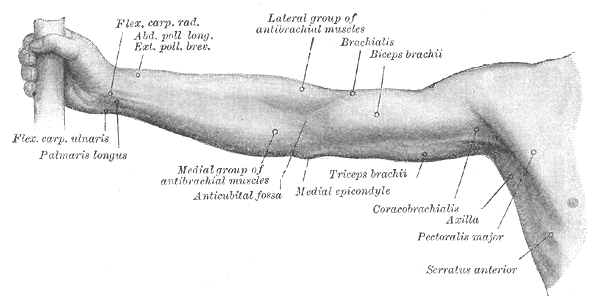
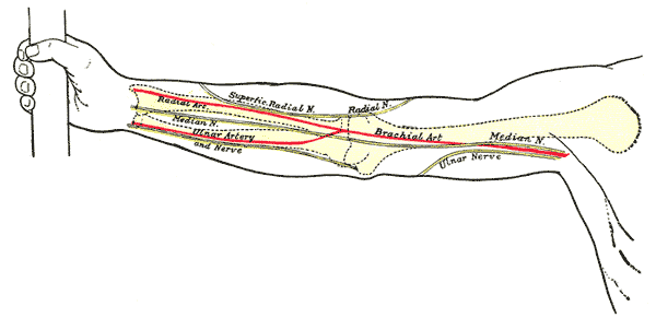
The cubital fossa (antecbuital fossa) is a inverted triangular shaped depression over the anterior aspect of the elbow joint which serves as a passageway for neurovascular structures passing between the upper arm and forearm.
Borders
The cubital fossa is triangular in shape and consists of three borders, a roof, and a floor:
- Medial border – lateral border of pronator teres
- Lateral border – medial border of brachioradialis
- Superior border – horizontal epicondylar line (drawn between the medial and lateral epicondyles of the humerus).
- Roof:
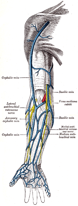
- deep fascia of the forearm reinforced medially by the bicipital aponeurosis
- medial cutaneous nerve of forearm; lateral cutaneous nerve of forearm
- basilic vein, cephalic vein, median cubital vein
- subcutaneous fat and skin
- Floor – brachialis (proximally) and supinator (distally)
Contents
Medial to lateral:
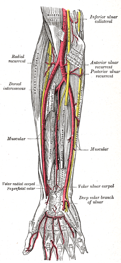
- Median nerve – travels medially through the cubital fossa, exiting by passing between the two heads of the pronator teres.
- It has a motor and sensory function in the anterior forearm and hand.
- Brachial artery – bifurcates into the radial and ulnar arteries at the apex of the cubital fossa.
- Biceps tendon – passes centrally through the cubital fossa and attaches the radial tuberosity (immediately distal to the radial neck).
- It gives rise to the bicipital aponeurosis which contributes to the roof of the cubital fossa.
- Radial nerve – travels along the lateral border of the cubital fossa and divides into superficial and deep branches.
- It has a motor and sensory function in the posterior forearm and hand.