

Overview
The median nerve is a major peripheral nerve of the upper limb, originating from the brachial plexus and providing motor and sensory innervation to parts of the forearm and hand.

Origin
Originates from the medial and lateral cords of the brachial plexus in the axilla
- Contains nerve roots from C6 – T1
Termination
Terminates in recurrent branch and palmar digital branch at hand

Course
- Enters arm from axilla at inferior margin of teres major
- Descends initially lateral to the brachial artery, then crosses anteriorly to course medial to the artery halfway down the arm.
- Enters anterior compartment of forearm via the cubital fossa.
- Passes between two heads of pronator teres, piercing the belly of the pronator teres, deep to bicipital aponeurosis and superficial to brachialis.
- Travels with median artery between flexor digitorum profundus and flexor digitorum superficialis.
- Gives off two major branches (anterior interosseous and palmar cutaneous)
- Finally enters hand via the carpal tunnel where it terminates as a recurrent branch and palmar digital branch.

Branches
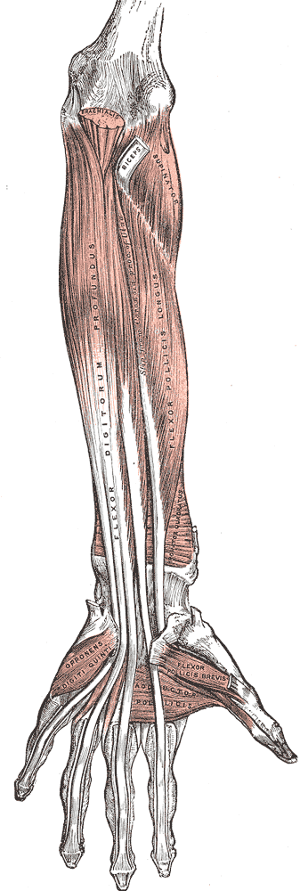
Forearm
- Anterior interosseous nerve – deep muscles of anterior forearm
- Flexor pollicis longus –
- Pronator quadratus –
- The radial (lateral) half of flexor digitorum profundus (inserting on the second and third digits)
- Palmar cutaneous nerve – skin of the lateral palm
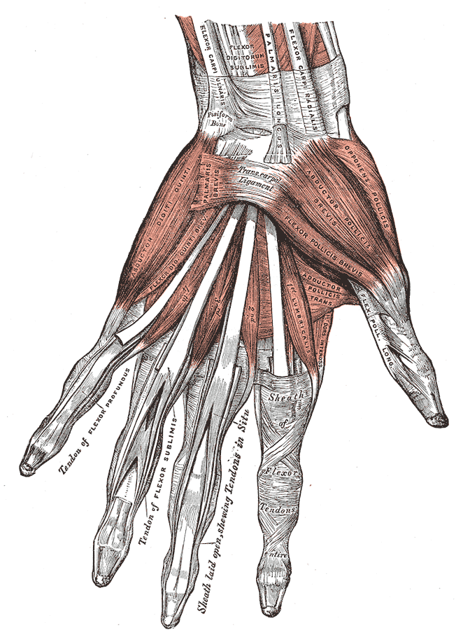
Hand
- Recurrent branch – thenar muscles (abductor pollicis, adductor pollicis, opponens pollicis, and flexor pollicis brevis)
- Palmar digital branch – palmar surface and fingertips of lateral three and a half digits, lateral two lumbrical muscles
Motor Function
In the forearm, the median nerve directly innervates the superficial and pronator flexor muscle groups:
Superficial compartment
- Pronator teres
- Origin: It has two origins, one from the medial epicondyle of the humerus, and the other from the coronoid process of the ulna.
- Insertion: Attaches laterally to the mid-shaft of the radius
- Actions: Pronation of the forearm
- Flexor carpi radialis
- Origin: Medial epicondyle of the humerus
- Insertion: Base of 2nd and 3rd metacarpals
Actions: Flexion and abduction at the wrist
- Palmaris longus
- Origin: Medial epicondyle of the humerus
- Insertion: Flexor retinaculum of the wrist
- Actions: Flexion at the wrist.
The fourth superficial flexor muscle, the flexor carpi ulnaris, is innervated by the ulnar nerve
Intermediate compartment
- Flexor digitorum superficialis
- Origin: It has two heads – one originates from the medial epicondyle of the humerus, the other from the radius
- Insertion: The muscle splits into four tendons at the wrist, which travel through the carpal tunnel, and attaches to the middle phalanges of the four fingers
- Actions: Flexes the metacarpophalangeal joints and proximal interphalangeal joints at the 4 finger. Flexes at the wrist.
The anterior interosseous nerve supplies the deep flexors:
Deep compartment
- Flexor pollicis longus
- Origin: Anterior surface of the radius and surrounding interosseous membrane.
- Insertion: Base of the distal phalanx of the thumb.
- Actions: Flexes the interphalangeal joint and metacarpophalangeal joint of the thumb.
- Innervation: Median nerve (anterior interosseous branch).
- Pronator quadratus
- Lateral half of the flexor digitorum profundus (the medial half of the muscle is innervated by the ulnar nerve, i.e. the little finger and ring finger).
- Origin: Ulna and associated interosseous membrane.
- Insertion: Splits into four tendons at the wrist, that pass through the carpal tunnel and attach to the distal phalanges of the four fingers.
- Actions: It is the only muscle that can flex the distal interphalangeal joints of the fingers. It also flexes at metacarpophalangeal joints and at the wrist.
Hand
The median nerve innervates some of the muscles in the hand via two branches.
The recurrent branch of the median nerve innervates the thenar muscles – muscles associated with movements of the thumb. The palmar digital branch innervates the lateral two lumbricals – these muscles perform flexion at the metacarpophalangeal joints and extension at the interphalangeal joints of the index and middle fingers
Sensory Functions
The median nerve is responsible for the cutaneous innervation of part of the hand. This is achieved via two branches:
Palmar cutaneous branch – arises in the forearm and travels into the hand. It innervates the lateral aspect of the palm. This nerve does not pass through the carpal tunnel, and is spared in carpal tunnel syndrome.
Palmar digital cutaneous branch – arises in the hand. Innervates the palmar surface and fingertips of the lateral three and half digits.
Relations
- 1st half of upper arm: lateral to brachial artery
- 2nd half of upper arm: medial brachial artery
- Cubital fossa: medial to brachial artery
- Hand: within carpal tunnel, deep to flexor retinaculum
Variants
- Bifid median nerve or high division
- Median nerve accompanied by persistent median artery of the forearm
- Accessory branches arising proximal or distal to the carpal tunnel
- Variant course of thenar branch