

Description
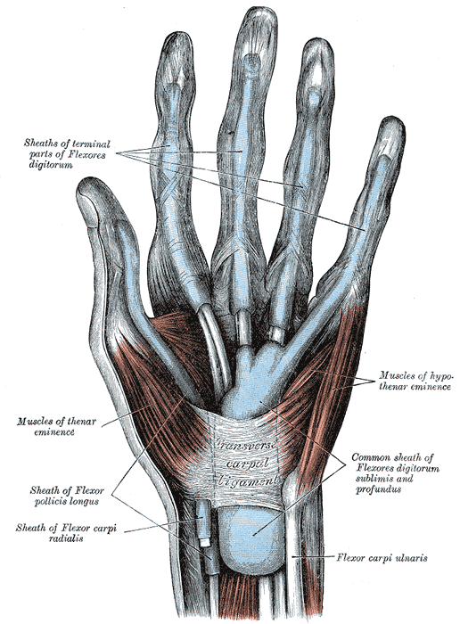
A fibro-osseous canal on the palmar side of the wrist that serves as a passageway for tendinous structures and the median nerve between the forearm and anterior hand. It lies between the flexor retinaculum and the carpal bones.

Borders
- Roof (superficial): Flexor retinaculum
- Thick fibrous band measuring 2-3 cm
- Attaches from the hook of hamate and pisiform medially
- Attaches to the tubercle of the scaphoid and the ridge of the trapezium laterally
- Proximal edge of the band corresponds to the distal dominant skin crease on the anterior wrist
- Contains the flexor carpi radialis tendon (not located within the carpal tunnel itself)
- Medial (ulnar border): lateral surface of hamate
- Lateral (radial border): medial surface of trapezium
- Floor (deep): carpal groove formed by the palmar aspect of the carpal bones
- hamate, pisiform, scaphoid, trapezium
Contents
Tendons
- Tendon of flexor pollicis longus (FPL)
- Most lateral structure
- Surrounded by own synovial sheath
- Runs in a groove on the trapezium
- FPL flexes the interphalangeal joint of the distal phalanx of the thumb
- Four tendons of flexor digitorum superficialis (FDS)
- Lie proximal to FDP in a row
- FDS flexes the middle phalanges of the four fingers at the proximal interphalangeal joint
- Four tendons of flexor digitorum profundus (FDP)
- Lie deep to FDS on one plane
- FDP flexes hand and both interphalangeal joints
- The eight tendons of the FDS and FDP share a common sheath, which does not invest them completely.
- Divides into recurrent branch and palmar digital branch
- Recurrent branch – thenar muscles (abductor pollicis, opponens pollicis, superficial head of the flexor pollicis brevis)
- Palmar digital branch – palmar surface and fingertips of lateral three and a half digits, lateral two lumbrical muscles
Relations
- Medially: Guyon’s canal
Variants
Nerve anomalies
- High bifurcation of median nerve
- Bifid median nerve
- Aberrant origin of motor branch of median nerve
- Transligamentous palmar cutaneous branch of median nerve
- Anomalous course of ulnar nerve
- Ulnar-to-median nerve connection (Marinacci communication)
Tendon anomalies
- Conjoint FPL and FDP II (Linburg-Comstock syndrome)
Vascular anomalies
- Persistent median artery
- Superficial ulnar artery
Muscle anomalies
- Presence of palmaris longus tendon within carpal tunnel
- Index lumbrical
- Flexor digitorum superficialis indicis