

Description
The ulnar nerve is a major peripheral nerve of the upper limb, arising from the medial cord of the brachial plexus (C8-T1).
The ulnar nerve runs down the medial arm and passes behind the medial epicondyle at the elbow, making it vulnerable to injury (cubital tunnel syndrome), then travels along the forearm into the hand. It supplies motor fibres to forearm muscles like the flexor carpi ulnaris and the medial half of the flexor digitorum profundus, and most of the intrinsic hand muscles. Its sensory distribution covers the medial hand, including the little finger and adjacent half of the ring finger.
Origin
Arises within the axilla region from the medial cord of the brachial plexus (spinal roots C8 – T1).

Course
- From the brachial plexus within the axilla region, it descends between the axillary artery (laterally) and vein (medially)
- It continues down on the medial aspect of the arm with the brachial artery laterally
- At the mid-point of the arm, it penetrates the medial fascial septum to enter the posterior compartment
- It passes posterior to the elbow through the cubital tunnel (between medial epicondyle and olecranon)
- In the forearm, it pierces the two heads of flexor carpi ulnaris, travelling deep to the muscle.
- It gives off two muscular branches, the palmar cutaneous branch and dorsal cutaneous branch
- At the wrist, the ulnar nerve travels superficially to the flexor retinaculum and medial to ulnar artery.
- It enters the hand via the ulnar canal (Guyon’s Canal)
Branches & Supply
Motor
Arm
- Flexor carpi ulnaris
- Flexes and adducts hand at the wrist
- Medial half of flexor digitorum profundus
- Flexes 4th and 5th digits at the distal interphalngeal nerves
(Note: all other muscles of the anterior forearm are innervated by median nerve)
Hand
- Intrinsic muscles of hand (except thenar muscle and lateral lumbricals which are supplied by median nerve)
- Hypothenar muscles
- Medial two lumbricals
- Adductor pollicis
- Palmar and dorsal interossei
- Palmar brevis
Sensory
Arm
- Palmar cutaenous branch – medial half of palm
- Dorsal cutaneous branch – dorsal surface of medial one and half fingers
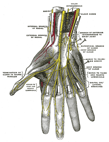
Hand
- Superficial branch – palmar surface of the medial one and half fingers
Normal Variants of the Ulnar Nerve
Motor–Sensory Communications
Martin-Gruber anastomosis
- Communication from the median nerve to the ulnar nerve in the proximal forearm
- Carries motor fibres, typically to intrinsic hand muscles
- Present in up to 15–30% of individuals
- Can cause confusing findings on nerve conduction studies
Marinacci anastomosis (Reverse Martin-Gruber)
- Communication from the ulnar nerve to the median nerve in the forearm
- Less common than Martin-Gruber
- Also affects interpretation of electrodiagnostic testing
Riche-Cannieu anastomosis
- Communication between the deep branch of the ulnar nerve and the recurrent branch of the median nerve in the thenar eminence
- Purely motor
- Can explain preserved thenar muscle function despite median nerve injury
Sensory Communications
Berrettini anastomosis
- Sensory communication between common digital branches of the median and ulnar nerves in the palm
- Found in 60–90% of individuals
- Can alter expected sensory distribution in the hand
Structural Variants
High division of the ulnar nerve
- Sensory and motor branches separate more proximally than usual, sometimes in the mid-forearm
- May mimic a bifid nerve on imaging or during dissection
Bifid ulnar nerve
- True duplication of the nerve trunk, typically in the forearm or cubital tunnel
- Can be mistaken for a tumour or schwannoma on imaging
- May share or duplicate vascular structures
Accessory ulnar nerve
- Extra fascicle or nerve branch running independently or in parallel to the main ulnar nerve
- May be a cause of recurrent symptoms if missed during surgical decompression
Intramuscular course
- Ulnar nerve passes through the substance of flexor carpi ulnaris or medial head of triceps
- Can predispose to dynamic compression with muscle hypertrophy or overuse
Relations
- The posterior bundle of the ulnar collateral ligament is deep to the ulnar nerve
Imaging Anatomy
US
- The ulnar nerve is visible as speckled or honeycomb in appearance from hypoechoic nerve fascicles and hyperechoic connective tissue
- Posterior to the medial epicondyle often appears hypoechoic from physiologic oedema but should not be enlarged