

Overview
A set of twelve paired flat bones which form a protective cage of the thorax.
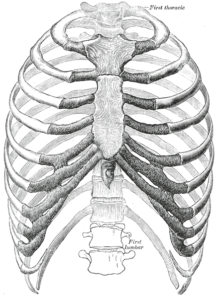
[foogallery id=”5420″]
Types (Typical & Atypical)
Typical Ribs
- Ribs 3 to 9 are typical ribs and the major landmarks are the head, neck, tubercle and body.
Atypical Ribs
- Ribs 1, 2, 10 11 and 12 are atypical ribs – they have features that are not common to all the ribs.
- Rib 1 is shorter and wider than the other ribs. Read more…
- Rib 2 is thinner and longer than rib 1, and has two articular facets at the head. It has a roughened area on its upper surface, from which the serratus anterior muscle originates.
- Rib 10 has one facet – for articulation with its numerically corresponding vertebrae.
- Ribs 11 and 12 have no neck and only contain one facet, which is for articulation with their corresponding vertebrae.
[foogallery id=”5415″]

Structure
- The head is wedge-shaped, bevelled by two articular facets which form synovial joints with the vertebral column posteriorly
- The upper facet articulates with the vertebra above
- The lower facet is vertical and articulates with its own vertebra
- The neck is flattened, with a sharp crest forming the upper border. The two laminae of the superior costotransverse ligament are attached to the crest
- The tubercle projects posteriorly from the lateral end of the neck. It has two facets:
- The smooth medial (articular) facet makes a synovial joint with the transverse process of its own vertebra
- The rough lateral (non-articular) facet gives attachment to the lateral costotransverse ligament
- The shaft slopes down and posteriorly to the angle of the rib, before arching anteriorly
- The diameter of the arch progressively increases with the inferior ribs
- The costal groove is located on the internal surface of the shaft, along its inferior border. It protects the passage of the neurovascular bundle to the thorax
- The neurovascular bundle, located in the costal groove between the internal intercostal muscle and innermost intercostal muscle, supplies most of the innervation and vascular supply to the thoracic wall. The structures include (from superior to inferior):
- Intercostal Vein –
- Intercostal Artery – The six-upper anterior intercostal arteries originate from the internal thoracic artery, and the lower five from the musculophrenic artery, itself a branch of the internal thoracic artery. Anterior intercostal arteries form anastomoses with the posterior intercostal arteries.
- Intercostal Nerve – originate segmentally from the ventral rami of T1 – T11. Supplies motor innervation to the intercostal muscles.
Articulations
Posterior articulations
All twelve ribs articulate posteriorly with the vertebrae of the spine. Each rib forms two joints:
- Costotransverse joint – Between the tubercle of the rib, and the transverse costal facet of the corresponding vertebrae.
- Costovertebral joint – Between the head of the rib, superior costal facet of the corresponding vertebrae, and the inferior costal facet of the vertebrae above.
Anterior articulations
- Ribs 1 – 7 attach independently to the sternum.
- Ribs 8 – 10 attach to the costal cartilages superior to them.
- Ribs 11 and 12 ‘float’ – i.e. they do not have an anterior attachment and end in the abdominal musculature.
Muscle Attachments
Intercostal muscle layers (from superficial to deep):
- External intercostal muscle
- Extend from lateral border of costal groove to the superior margin of the ribs below
- Fibres course obliquely in an anteroinferior direction
- Elevate the ribs in inspiration
- Internal intercostal muscle
- Extend from inferior edge of costal groove to superior margin of the ribs below
- Fibres course obliquely in a posteroinferior direction
- Depress the ribs in forced expiration
- Innermost intercostal muscle
- Extend in same direction as internal intercostal muscle.
- Fibres extend from the medial edge of the costal groove to the medial surface of the rib below
- Neurovascular bundle passes through opening between internal and innermost layers
- Depress the ribs in forced expiration
Subcostales
- located within the same plane as the innermost intercostals.
- span one or multiple ribs and become more numerous within the inferior regions of the posterior thoracic wall. They extend from the inner surface of one rib to the inner surface of either the next rib or even the one below that. This muscle assists the internal intercostal muscles.
Transversus thoracis
These muscles are found deep within the anterior thoracic wall. The transversus thoracic muscles originate from the posterior surface of the xiphoid process and the lower part of the body of the sternum. The fibres pass superolaterally to insert into the internal surface of costal cartilages of ribs two to six. This muscle assists in depression of the ribs.
Serratus posterior
This muscle is present posteriorly within the thoracic wall. It is split into superior and inferior fibres. The inferior fibres originate from the spinous processes of the T11 to L2 vertebrae and attach to the lower borders of ribs eight to twelve near the angle. The part of the muscle is thought to depress the ribs. The superior fibres originate from the spinous processes of the C7 to T3 vertebrae and attach to the superior borders of ribs two to four. This part of the muscle is thought to elevate the ribs.
Levatores costarum
Levatores costarum originates from the transverse processes of vertebrae C7 to T11 and attaches to the external surface between the tubercle and the angle of the rib below. Its action is to assist in elevation of the ribs.
Variants
- Cervical rib
- Intrathorcic rib
- Lumbar and pelvic ribs
- Forked rib
- Fusion and bridging
- Pseudarthrosis of the first rib
First Rib |
|---|
| CATEGORY 1UNSPECIFIED |
Overview
The first rib is the strongest, broadest, flattest and most curved rib

Surfaces
- Superior surface
- Forms part of the root of the neck
- Grooved obliquely at its greatest lateral convexity for the subclavian artery and the lower trunk of the brachial plexus (behind the artery)
- Inferior (pleural) surface
- Forms part of the roof of the thoracic cavity
Components
- The head is small and has a single facet that forms a synovial joint with the upper part of the body of T1 vertebra
- The neck slopes upwards, backwards and laterally from the head to the tubercle
- The tubercle is the most posterior and highest part of the rib. It projects from the inner border, anterior to the subclavian groove
- The subclavian groove
- The groove for the subclavian vein
- The shaft expands anteriorly into a concavity for the first costal cartilage.
Relations
The sympathetic trunk (or cervicothoracic ganglion) lies in contact with the anterior border of the neck alongside the head.
Lateral to it the first posterior intercostal vein and then the superior intercostal artery lie in contact, and more lateral still the first thoracic nerve lies in front of the neck.
The cervical dome of the pleura and the apex of the lung hold these vessels and nerves against the front of the neck of the rib.
The rib broadens at the junction of neck and shaft, where a prominent tubercle projects back to form the most posterior convexity of the rib. It is a fusion of tubercle and angle. Medially it has a convex facet for a corresponding concavity on the first transverse process. The lateral prominent part of the tubercle receives the lateral costotransverse ligament. The undersurface of the shaft, crossed obliquely by the small first intercostal nerve and vessels, is covered by
adherent parietal pleura. The external and internal intercostal muscles are attached together to the outer rim of
this surface. The outer border is blunt between the tubercle and subclavian groove and the first digitation of
serratus anterior is attached to the anterior part of this blunt area. The concave internal border of the shaft gives
attachment to the suprapleural membrane in front of the subclavian groove (Fig. 4.5).
The fibres in contact with the rib are all T1, the C8 fibres lying above them (Fig. 4.34).
Muscle Attachments
- Scalenus medius – attaches to large quadrangular area of supper surface, between subclavian groove and tubercle
- Scalenus anterior – attaches to the tubercle via its tendon and extends along a traingular impression on the upper surface
- Anterior to the attachment of scalenus anterior is another groove on the upper surface of the shaft in which the subclavian vein lies. The anterior end of the shaft expands into a concavity for the first costal cartilage. From the upper surface of this junction subclavius arises from both bone and cartilage.
https://pubs.rsna.org/doi/pdf/10.1148/radiographics.19.5.g99se011125#:~:text=Normal%20rib%20variants%20include%20cervical,the%20radiologist%20to%20other%20injuries.