

Nil
Category 2
• Galea
• Blood supply to scalp
Category 3
• Frontalis muscle
• Occipitalis muscle
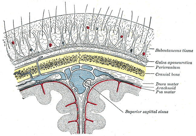

The scalp extends from the supraorbital margins anteriorly to the highest nuchal lines.
Layers
Skin – thin, contains sweat and sebaceous glands and hair follicles
Connective tissue (dense) – thick, richly vascularised subcutaneous layer, supplied with cutaneous nerves
Aponeurosis (galeal/epicranial) – broad, strong tendinous sheet, serves as an attachment for muscles of the cranium
Loose connective/areolar tissue – sponge-like layer, potential space
Pericranium – dense layer of connective tissue, external periosteum of the neurocranium
Galea Aponeurotica |
|---|
| CATEGORY 2PENDING |
Also known as the epicranial aponeurosis, is a tough layer of dense fibrous connective tissue that covers the upper part of the cranium and serves as an intermediate tendon between the frontal and occipital belly of occipitofrontalis muscle.
Attachments
- anteriorly – frontalis
- posteriorly:
- occipitalis
- external occipital protuberance
- highest nuchal lines of the occipital bone
- laterally – anterior and superior auricular muscles
Blood Supply |
|---|
| CATEGORY 2PENDING |
- Occipital artery – emerges from the apex of posterior triangle
- Superficial temporal artery
- Supraorbital artery – supplies front of scalp up to vertex, branch of the internal carotid
- Supratrochlear artery – branch of the internal carotid.
Innervation
- Anterior to the auricles of the external ear – branches of all three divisions of the trigeminal nerve (CN V)
- Posterior to the auricles of the external ear – spinal cutaneous nerves of C2 and C3
Lymph drainage
- No lymph nodes within the scalp
- Posterior half of scalp drains to occipital and mastoid nodes
- Anterior half of scalp drains to preauricular nodes
Occipitofrontalis muscle |
|---|
| CATEGORY 2PENDING |
A muscle which covers parts of the skull, consisting of two parts; the frontal and the occipital belly. Its primary function is for facial expressions (draws the scalp back to raise eyebrows and wrinkle the forehead).
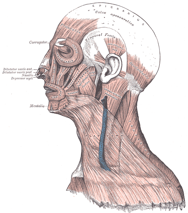
Frontal Belly
- Blood supply: Supraorbital and supratrochlear arteries
- Origin: galea aponeurotica
- Inserts: fascia of the fascial muscles and skin above the eyes and nose
- Innervation: facial nerve
Occipital Belly
- Origin: lateral two-thirds of the highest nuchal line, the mastoid process of the temporal bone
- Inserts: galea aponeurotica
- Blood supply: occipital artery
- Innervation: facial nerve