

Overview
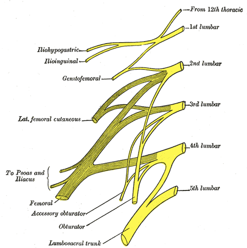
The femoral nerve is one of the major peripheral nerves of the lower limb and the largest nerve of the lumbar plexus
Origin
Anterior rami of lumbar roots L2, L3, L4 of lumbar plexus

Course & Relations
- From the lumbar plexus, the femoral nerve travels inferiorly through the psoas major
- It courses between the iliacus (posteriorly) and psoas major (anteriorly) muscles, supplying motor branches to both.
- The femoral nerve then passes underneath the inguinal ligament to enter the femoral triangle
- Contents of the femoral triangle (lateral to medial): femoral nerve, femoral artery, femoral vein, femoral canal, and lymphatic vessels
- Approximately 4cm below the inguinal ligament, the femoral nerve is divided into anterior and posterior divisions by the lateral femoral circumflex artery.
Termination
- The terminal cutaneous branch of the femoral nerve is the saphenous nerve which innervates the medial aspect of the leg and the foot.
- It travels through the adductor canal (accompanied by the femoral artery and vein) and exits prior to the adductor hiatus.
Divisions
Anterior Division
- Four terminal branches (two motor and two sensory)
- Motor: Nerve to pectineus, nerve to sartorius
- Sensory: Medial cutaneous nerve of the thigh and the intermediate cutaneous nerve. These nerves are responsible for the anteromedial sensory innervation of the thigh.
Posterior Division
- One sensory nerve, four motor branches
- Sensory:
- Saphenous nerve – the largest cutaneous branch of the femoral nerve, responsible for sensory innervation along the anteromedial and posteromedial aspect of the leg, into the medial foot.
- The femoral nerve becomes the saphenous nerve when it passes through the adductor canal. It continues to travel along the medial aspect of the tibia until about halfway down the tibia, where it finally divides into two branches. One branch is more posterior and ends at the ankle. The other branch is more anterior and continues along the medial aspect of the foot and terminates at the hallux.
- Motor:
- Nerves to the rectus femoris, vastus medialis, lateralis, and intermedius muscles (muscles of the anterior compartment of the thigh)
- Articular joint nerves – branches which supply the knee and hip joints innervating the fibrous capsule, ligaments, and synovial membranes of the joints
Motor Functions
- Hip flexors:
- Pectineus – adducts and flexes the thigh, assists with medial rotation of the thigh.
- Iliacus – acts with psoas major and psoas minor (forming iliopsoas) to flex the thigh at the hip joint and stabilise the hip joint.
- Sartorius – flexes, abducts and laterally rotates the thigh at the hip joint. Flexes the leg at the knee joint.
- Knee extensors:
- Quadriceps femoris (rectus femoris, vastus lateralis, vastus medialis and vastus intermedius) – extends the leg at the knee joint. Rectus femoris also steadies the hip joint and assists iliopsoas in flexing the thigh.
Sensory Functions
- Anterior cutaneous branches – derived from the anterior division of the femoral nerve. They supply the skin of the anteromedial thigh.
- Saphenous nerve – a continuation of the posterior division of the femoral nerve. It supplies the skin of the medial leg and foot.
Variant anatomy
- Femoral nerve splits into two or three separate slips within the psoas major but unites to descend as a single bundle
- Accessory femoral nerve: fibers arise separately in lumbar plexus, passes anterior to femoral nerve, may terminate as saphenous nerve / cutaneous branch
- femoral branch replacing lateral femoral cutaneous nerve which usually branches directly off lumbar plexus
- saphenous nerve terminating at knee with distribution replaced by branch of the tibial nerv