

Overview
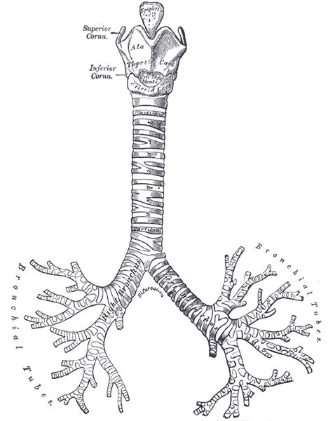
The trachea is a cartilaginous tube that conducts air from the upper respiratory tract to the lungs via the tracheobronchial tree. It is a C-shaped structure, composed of hyaline cartilage rings on the anterior and lateral walls, with the trachealis smooth muscle forming the posterior border of the trachea.
Origin
The inferior margin of the cricothyroid cartilage at level of C6
Course
- Descends through the midline of the anterior neck, anterior to the oesophagus
- It courses posteriorly to enter the superior mediastinum through the superior thoracic aperture
- The angle of descent becomes more horizontal with age due to kyphotic changes in the spine and tethering of the left main stem bronchus under the aortic arch

Termination
The carina – a bifurcation of the trachea into left and right main bronchi at level of T4-5 posteriorly or sternal angle (of Louis)
- The vertical position in the mediastinum changes with phases of respiration
Structure
- Consist of 15 – 20 C-shaped ‘rings’ composed of hyaline cartilage, oriented such that the C opens posteriorly
- Each ring is interconnected by the annular ligament
- A membranous wall connects the arms of the C-shaped cartilage rings posteriorly
- The trachealis muscle runs longitduinally on the posterior aspect of the membranous wall, abutting the anterior oesophagus
- Functionally aids in expectoration with contraction against a closed epiglottis, where a sudden opening results in forceful explusion.
- Lowest tracheal ring has an inferior hook, projecting posteriorly in the midline between main bronchi origins (carina)
Blood supply
Cervical trachea
- The tracheoesophageal branches of the inferior thyroid arteries supplies the cervical trachea from the right and left thyrocervical trunks that branch off the subclavian arteries
- The first tracheoesophageal branch supplies the lower cervical trachea, the second branch supplies the middle cervical trachea and the third branch supplies the upper cervical trachea
- The superior thyroid artery does not directly supply the trachea but forms an anastomosis with the inferior thyroid artery where fine branches supply the thyroid isthmus and the adjacent anterior tracheal wall.
Thoracic trachea
- The thoracic trachea and carina receive blood from the bronchial arteries (superior, middle and inferior) arising directly from the aorta
- The superior bronchial artery comes off the anteromedial aspect of the descending thoracic aorta lateral to the carina and posterior to the left main bronchus.
- The anterior branch courses over the proximal left main stem bronchus, which it supplies on its way to providing blood to the anterior portion of the carina
- The principal and posterior branches pass behind the posterior wall of the oesophagus to supply the proximal right main stem bronchus
- These branches may also arise from an intercostal bronchial trunk
- The middle bronchial artery comes off the aorta and travels posterior to the medial aspect of the left main stem bronchus to supply the carina as it anastomoses with the anterior branch of the superior bronchial artery or higher tracheoesophageal branches
- The inferior bronchial artery arises off the right posteromedial ascending thoracic aorta to supply the left main stem bronchus
Venous drainage
- Trachea drained by inferior thyroid veins
- Left bronchial veins into left superior intercostal vein or accessory hemiazygos vein
- Right bronchial veins into right superior intercostal vein or Azygos vein
Lymphatic
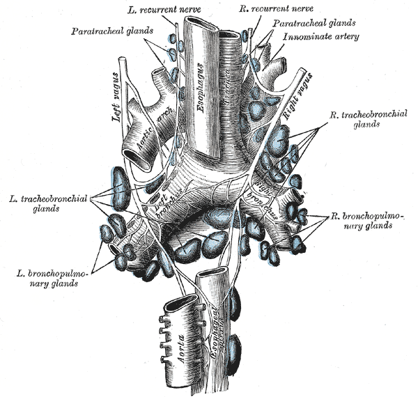
Lymphatic drainage of the trachea is made up of pretracheal, paratracheal, and inferior deep cervical nodes. The lymphatic drainage path from distal to proximal in the lung is as follows: pulmonary and intrapulmonary nodes; bronchopulmonary and hilar nodes; inferior tracheobronchial (carina) and superior tracheobronchial nodes; paratracheal nodes; inferior deep cervical nodes. Some drainage will also occur through the aortic node.
Pretracheal and paratracheal lymph nodes into bronchomediastinal trunk
Innervation
- Vagus and recurrent laryngeal nerves
- Sympathetic fibers from upper ganglia of sympathetic trunks
Relations
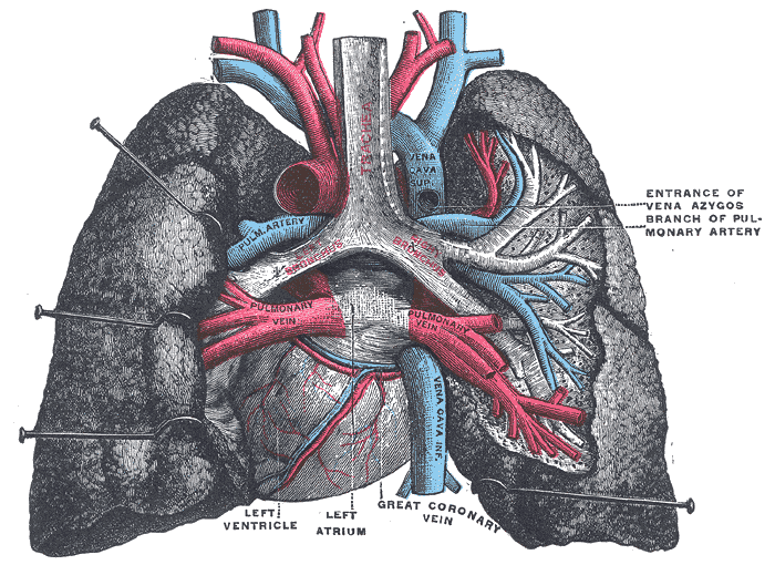
- Anterior
- Strap muscles
- Thyroid isthmus – crosses the anterior trachea at the 2nd or 3rd tracheal ring
- Thyroid vessels
- Thoracic branches of vagus nerve
- Thymus
- Sternum
- Left brachiocephalic veins
- Left Brachiocephalic artery
- Left common carotid arteries.
- Posterior
- Lateral
- Carotid sheath
- Lateral lobes of thyroid (anterolateral)
- Recurrent laryngeal nerve (posterolateral)
- Azygos arch (right)
- Arch of aorta (left)
- Vagus nerve
- Lymph nodes
- Lung and pleura
- Superior
- Cricoid cartilage
- Inferior
- Pulmonary trunk
Anatomical Variants
- Tracheal bronchus (pig bronchus) – accessory bronchus originates from the supracarinal trachea
- Oesophageal bronchus – communicating bronchopulmonary foregut malformation (bronchus arises directly from oesophagus)
- Additional bronchus from bronchus intermedius
- Supernumerary segmental bronchi
- Tracheal diverticulum
- Paratracheal air cyst
- Short trachea
- Lunate trachea – lunate shaped, rather than round/oval
- Bronchial atresia
https://www.ncbi.nlm.nih.gov/pmc/articles/PMC5900092/